Download
1 / 54
550 likes | 921 Views
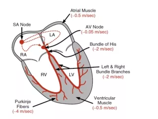
Cardiac Arrhythmias. Atrial Depolarization and the Inscription of the P-wave. Ventricular Depolarization and the Inscription of the QRS complex. Ventricular Repolarization and the Inscription of the T-wave. The ECG Complex with Interval and Segment Measurements.

E N D
Ventricular Depolarization and the Inscription of the QRS complex
Ventricular Repolarization and the Inscription of the T-wave
The Concept of a “Lead” Summary of the “Limb Leads” LEAD AVR LEAD AVL -150o -30o Each of the limb leads (I, II, III, AVR, AVL, AVF) can be assigned an angle of clockwise or counterclockwise rotation to describe its position in the frontal plane. Downward rotation from 0 is positive and upward rotation from 0 is negative. 0o LEAD I 60o 120o LEAD II 90o LEAD III LEAD AVF
The “Precordial Leads” 4th intercostal space V1 V2 Each of the 6 precordial leads is unipolar (1 electrode constitutes a lead) and is designed to view the electrical activity of the heart in the horizontal or transverseplane V3 V6 V5 V4 V1 - 4th intercostal space - right margin of sternum V2 - 4th intercostal space - left margin of sternum V3 - linear midpoint between V2 and V4 V4 - 5th intercostal space at the mid clavicular line V5 - horizontally adjacent to V4 at anterior axillary line V6 - horizontally adjacent to V5 at mid-axillary line
Diagnosis and treatment of arrhythmias can be simplified by using the following checklist when looking at an electrocardiographic display: • 1. What is the heart rate? • 2. Is the rhythm regular? • 3. Is there one P wave for each QRS Complex? • 4. Is the QRS complex normal? • 5. Is the rhythm dangerous? • 6. Does the rhythm require treatment?
Normal Sinus Rhythm www.uptodate.com Implies normal sequence of conduction, originating in the sinus node and proceeding to the ventricles via the AV node and His-Purkinje system. EKG Characteristics: Regular narrow-complex rhythm Rate 60-100 bpm Each QRS complex is proceeded by a P wave P wave is upright in lead II & downgoing in lead aVR
Sinus Bradycardia • HR< 60 bpm; every QRS narrow, preceded by p wave • Can be normal in well-conditioned athletes • HR can be<30 bpm in children, young adults during sleep, with up to 2 sec pauses
Sinus bradycardia--etiologies • Normal aging • 15-25% Acute MI, esp. affecting inferior wall • Hypothyroidism, infiltrative diseases (sarcoid, amyloid) • Hypothermia, hypokalemia • SLE, collagen vasc diseases • Situational: micturation, coughing • Drugs: beta-blockers, digitalis, calcium channel blockers, amiodarone, cimetidine, lithium
Sinus bradycardia--treatment • No treatment if asymptomatic • Sxs include chest pain (from coronary hypoperfusion), syncope, dizziness • Office: Evaluate medicine regimen—stop all drugs that may cause • ATROPINE 0.5 mg (max dose 0.04 mg/kg) • Ephedrine 5-25 mg • Dopamine 5-20 microgram/kg/min • Epinephrine 2-10 microgram/min
Sinus tachycardia • HR > 100 bpm, regular • Often difficult to distinguish p and t waves
Fever Hyperthyroidism Effective volume depletion Anxiety Pheochromocytoma Sepsis Anemia Exposure to stimulants (nicotine, caffeine) or illicit drugs Hypotension and shock Pulmonary embolism Acute coronary ischemia and myocardial infarction Heart failure Chronic pulmonary disease Hypoxia Sinus tachycardia--etiologies
Sinus Tachycardia--treatment • Office: evaluate/treat potential etiology :check TSH, CBC, optimize CHF or COPD regimen, evaluate recent OTC drugs • Verify it is sinus rhythm • If no etiology is found and is bothersome to patients, can treat with beta-blocker
Sinus Arrhythmia • Variations in the cycle lengths between p waves/ QRS complexes • Will often sound irregular on exam • Normal p waves, PR interval, normal, narrow QRS
Sinus arrhythmia • Usually respiratory--Increase in heart rate during inspiration • Exaggerated in children, young adults and athletes—decreases with age • Usually asymptomatic, no treatment or referral • Can be non-respiratory, often in normal or diseased heart, seen in digitalis toxicity • Referral may be necessary if not clearly respiratory, history of heart disease
Paroxysmal Supraventricular Tachycardia(PSVT) • Heart rate : 130-270 • Rhythm : regular • QRS : normal • P/QRS: 1 : 1 relationship, although the P wave may often be hidden in the QRS complex or T wave.
PSVT treatment Vagal maneuvers such as carotid sinus massage should be applied only to one side Adenosine, which is the drug of choice, is given by 6-mg rapid (2 seconds) intravenous bolus, preferably through an antecubital or central vein. If no response is elicited, second and third doses of 12 to 18 mg of adenosine may be administered by rapid intravenous bolus Verapamil(2.5 to 10 mg given intravenously) Amiodarone (150-mg infusion over a 10-minute period for the loading dose) is a recent addition.
Atrial Fibrillation • Irregular rhythm • Absence of definite p waves • Narrow QRS • Can be accompanied by rapid ventricular response
Hypertension Hyperthyroidism and subclinical hyperthyroidism CHF (10-30%), CAD Uncommon presentation of ACS Mitral and tricuspid valve disease Hypertrophic cardiomyopathy COPD OSA ETOH Caffeine Digitalis Familial Congenital (ASD) Atrial Fibrillation—causes and associations
Atrial fibrillation--assessment • H & P—assess heart rate, sxs of SOB, chest pain, edema (signs of failure) • If unstable, need to cardiovert • Echocardiogram to evaluate valvular and overall function • Check TSH • Assess onset of sxs—in the last 24-48 hours? Sudden onset? Or no sxs?
Atrial fibrillation--management • Rhythm vs Rate control—if onset is within last 24-48 hours, may be able to arrange cardioversion—use heparin around procedure • Need TEE if valvular disease (high risk of thrombus) • If unable to definitely conclude onset in last 24-48 hours: need 4-6 weeks of anticoagulation prior to cardioversion, and warfarin for 4-12 weeks after
Atrial fibrillation--management • β-Blockers such as esmolol (1 mg/kg by intravenous bolus) or propranolol • Calcium channel blockers such as verapamil (5 to 10 mg given intravenously) or diltiazem
Atrial fibrillation--management • Goal INR of 2.5 (2.0-3.0) • Rhythm control---second line approach, if unable to control rate or pt with persistent sxs • Can also consider radiofrequency ablation at pulm veins
If the ventricular response is excessively rapid or hemodynamic instability is present, or both, the following guidelines should be used • Synchronized DC cardioversionstarting at a relatively high energy of 100 J and gradually increasing to 360 J is indicated • The class III antiarrhythmic agentibutilide(Corvert, 1 mg in 10 mL saline or [D5W] infused slowly intravenously over a 10-minute period) has been documented to convert atrial flutter to sinus rhythm in most patients • Procainamide (5 to 10 mg/kg for the intravenous loading dose, infused no faster than 0.5 mg/kg/min) and amiodarone
PAC • P wave from another atrial focus • Occurs earlier in cycle • Different morphology of p wave
PAC • Benign, common cause of perceived irregular rhythm • Can cause sxs: “skipping” beats, palpitations • No treatment, reassurance • With sxs, may advise to stop smoking, decrease caffeine • Can use beta-blockers to reduce frequency
PVC • Extremely common throughout the population, both with and without heart disease • Usually asymptomatic, except rarely dizziness or fatigue in patients that have frequent PVCs and significant LV dysfunction
PVC • No treatment is necessary, risk outweighs benefit • Reassurance • Optimize cardiac and pulmonary disease management
PVC treatment • treatment is generally dictated by the presence of symptoms attributable to the VPBs. • correct any underlying abnormalities such as decreased serum potassium or low arterial oxygen tension. • lidocaine ; initial bolus dose of 1.5 mg/kg. Recurrent VPBs can be treated with a lidocaine infusion at 1 to 4 mg/min; • additional therapy includes esmolol, propranolol, procainamide, quinidine, disopyramide, atropine, verapamil, or overdrive pacing
Non-sustained Ventricular tachycardia • Defined as 3 or more consecutive ventricular beats • Rate of >120 bpm, lasting less than 30 seconds • May be discovered on Holter, or other exercise testing
Non-sustained ventricular tachycardia • Need to exclude heart disease with Echo and stress testing • If normal, there is no increased risk of death • May need anti-arrhythmia treatment if sxs • In presence of heart disease, increased risk of sudden death • Need referral for EPS and/or prolonged Holter monitoring
Ventricular tachycardia treatment • amiodaroneadministered as one or more intravenous doses of 150 mg in 100 mL saline or D5W over a period of 10 minutes, followed by an intravenous infusion of 1 mg/min for 6 hours and 0.5 mg/min • hypotension and bradycardia are its main side effects
Ventricular fibrillation • Cardiopulmonary resuscitation • DefibrillationAsynchronous external defibrillation should be performed with a DC defibrillator using incremental energies in the range of 200 to 360 J. • 1 g of magnesium sulfate may facilitate defibrillation
Assess and support ABC • Give oxygen • Monitor ECG , BP, pulse oximetry • Check unstable signs ; chest pain, hypotension -- unstable cardioversion • Stablish IV access • Obtain 12 lead ECG • Identify and treat reversible causes
Tachycardia with pulse • 1-ABC – oxygen – ECG monitor • 2-is patient stable? • 3-unstable IV access , sedation , cardioversion • 4- stable 12 LEAD ECG , IV access ,check QRS • 5-narrow QRS REGULAR (PSVT) VAGAL MANEUVRE , ADENOSINE • irregular (AF) control HR beta blocker , ca channel blocker
6-wide QRS regular (VT) amiodarone , cardioversion • Irregular AF with abberancy (AF + WPW) avoid verapamil , adenosine , digoxin , diltiazem
Bradycardia • Rate? 30 bpm • Regularity? regular • P waves? normal • PR interval? 0.12 s • QRS duration? 0.10 s Interpretation? Supraventricular Bradycardia
Supraventricular Bradyarrhythmia • sinus or junctional in origin • second-degree (types I and II) or third-degree atrioventricular (AV) block • Treatment is indicated whenever the bradycardia, regardless of type, leads to a significant decrease in systemic arterial pressure • Initial treatment is atropine, 0.5 to 1.0 mg intravenously and repeated as needed at 3- to 5-minute intervals up to 0.04 mg/kg.[126 • dopamine (5 to 20 µg/kg/min) or epinephrine (2 to 10 µg/min) • External transcutaneous pacing