Download
1 / 71
710 likes | 880 Views
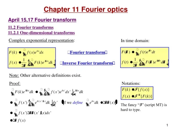
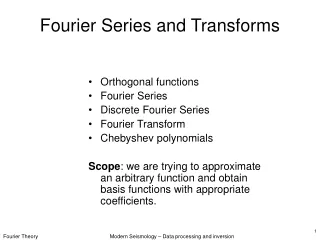
Fourier Transforms. Topics. I. Why you want one. II. What it tells you. III. How to think about them. IV. How to get one. I. Why you want one. A helical tube of virus head protein. The protein subunits can be seen clearly in some places but not others.

E N D
Topics I. Why you want one. II. What it tells you. III. How to think about them. IV. How to get one.
A helical tube of virus head protein. The protein subunits can be seen clearly in some places but not others. Although we see some regularities, they are not everywhere. Is this simply a bad image?
Photographic image superposition (averaging) by Roy Markham. The image is shifted and added to the original.
Superimposed images using Adobe Photoshop. I used Markham’s lattice to determine how much to shift by.
How would I figure out the distance and direction to shift if there weren’t a guide? 1. I could guess and pick the answer (image) that I liked best. 2. I could try all possible shifts and pick out the image with the strongest features (measured objectively rather than subjectively). The Fourier transform carries out the essence of method 2.
EM of catalase Optical diffraction pattern weak and strong exposure. (Erikson and Klug, 1971)
Bacterial rhodopsin in glucose Fourier transform of image.
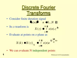
II. What the Fourier transform tells you.
What you see. What you get Spots Excited
What you see. What you get Spots Excited Spot positions Unit cell size and shape
What you see. What you get Spots Excited Spot positions Unit cell size and shape Spot size Size of coherent domains
What you see. What you get Spots Excited Spot positions Unit cell size and shape Spot size Size of coherent domains Intensity relative to background Signal/noise ratio
What you see. What you get Spots Excited Spot positions Unit cell size and shape Spot size Size of coherent domains Intensity relative to background Signal/noise ratio Distance to farthest spot Resolution
What you see. What you get Spots Excited Spot positions Unit cell size and shape Spot size Size of coherent domains Intensity relative to background Signal/noise ratio Distance to farthest spot Resolution Amplitude and phases of spots Structure of molecules
What you see. What you get Spots Excited Spot positions Unit cell size and shape Spot size Size of coherent domains Intensity relative to background Signal/noise ratio Distance to farthest spot Resolution Amplitude and phases of spots Structure of molecules Positions of Thon rings Amount of defocus
What you see. What you get Spots Excited Spot positions Unit cell size and shape Spot size Size of coherent domains Intensity relative to background Signal/noise ratio Distance to farthest spot Resolution Amplitude and phases of spots Structure of molecules Positions of Thon rings Amount of defocus Ellipticity of Thon rings Amount of astigmatism
What you see. What you get Spots Excited Spot positions Unit cell size and shape Spot size Size of coherent domains Intensity relative to background Signal/noise ratio Distance to farthest spot Resolution Amplitude and phases of spots Structure of molecules Positions of Thon rings Amount of defocus Ellipticity of Thon rings Amount of astigmatism Asymmetric intensity of Thon rings Amount of instability
What you see. What you get Spots Excited Spot positions Unit cell size and shape Spot size Size of coherent domains Intensity relative to background Signal/noise ratio Distance to farthest spot Resolution Amplitude and phases of spots Structure of molecules Positions of Thon rings Amount of defocus Ellipticity of Thon rings Amount of astigmatism Asymmetric intensity of Thon rings Amount of instability Direction of asymmetry Direction of instability
What you see. What you get Spots Excited Spot positions Unit cell size and shape Spot size Size of coherent domains Intensity relative to background Signal/noise ratio Distance to farthest spot Resolution Amplitude and phases of spots Structure of molecules Positions of Thon rings Amount of defocus Ellipticity of Thon rings Amount of astigmatism Asymmetric intensity of Thon rings Amount of instability Direction of asymmetry Direction of instability
III. How to think about Fourier transforms.
Some simple 1-D Fourier transforms
F(X)={sin(aX)/(X) The Fourier transform of a box. -a/2 a/2 X 1/a f(x)=1 if –a/2<x<a/2 f(x)=0 otherwise
0 X F(X)=(X) Fourier transform of a constant. x f(x)=1
+1/a -1/a F(X)=0.5[(X+1/a)+ (X-1/a)] Fourier transform of a cosine wave. | x=a X f(x)=cos(2x/a)
F(X)=a •exp(-X2a2) Fourier transform of a Gaussian. X a 1/a x f(x)=exp(-x2/a2)
0 1/a -3/a -2/a -1/a 2/a 3/a F(X)=…(X+1/a)+(X)+(X-1/a)… The Fourier transform of a lattice. 0 a -3a -2a -a 2a 3a x X f(x)=…(x+a)+(x)+(x-a)…
1/d F(X) In 2D the transform of a row of periodically placed points is a set of lines. This set of lines is perpendicular to the line joining the points. d f(x)
1/d F(X) In 3D, the transform of a row of points is a set of planes. The planes are perpendicular to the line joining the points. d f(x)
1/d F(X) In 3D, the transform of a plane of evenly spaced lines is a plane of evenly placed lines. These lines in real space are perpendicular to the plane containing the lines in reciprocal space (and vice versa). d f(x)
Some Rules for Fourier Transforms
1. Inverse Fourier transform: If F(X)=FT[f(x)], then f(x)=IFT[F(X)] where FT=Fourier transform & IFT=Inverse Fourier transform. USE: If you can obtain the Fourier transform, F(X), of an object, you can regenerate the object itself. This is the basis of x-ray crystallography and some 3D reconstruction algorithms.
2. Multiplication by a constant: FT[ a•f(x) ] = a•F(X) Special case: FT[ -f(x) ] = -F(X) = F(X)•ei USE: If you multiply the density by a constant, you multiply its Fourier transform by the same constant. If you reverse the contrast of an object, you get the same transform except the phases are changed by 180º (Babinet’s principle). Thus the phases obtained from images of negatively stained objects will differ by 180º from those of an ice-embedded object.
3. The addition of two density distributions (objects): FT[ f(x) + g(x)] = F(X) + G(X) USE: The Fourier transform of a heavy atom derivative is equal to the Fourier transform of the protein plus the Fourier transform of the constellation of heavy atoms. This allows one to use heavy atoms to determine the Fourier transform of the protein if the transform of the heavy atom constellation can be deduced.
4. The Fourier transform of a stretched object: FT[ f(ax) ] = F(x/a) USE: If you stretch/magnify an object by a factor of a, you squeeze/demagnify its transform by factor of a.
5. Rotation of an object: FT[ f{ x•cos(a) + y•sin(a), -x sin(a) + y •cos(a)}] = F { X•cos(a) + Y•sin(a), -X sin(a) + Y •cos(a)} USE: If you rotate an object by an angle a, you rotate its transform by the same angle.
6. Fourier transform of a shifted object: FT[ f(x-a) ] = F(X)•eiaX USE: If you shift an object by +a, you leave the amplitudes of its transform unchanged but its phases are increased by aX radians = 180ºaX degrees. The electron diffraction pattern is not sensitive to movement of the specimen since the intensities do not depend on phases. Vibration of the specimen does not affect the electron diffraction patterns as it does the images.
7. The section/projection theorem: FT[ f(x,y,z)dx ] = F(0,Y,Z) USE: The Fourier transform of a projection of a 3D object is equal to a central section of the 3D Fourier transform of the object. An electron micrograph is a projection of a 3D object. Its transform provides one slice of the 3D transform of the 3D object. By combining the transforms of different views, one builds up the 3D transform section by section. One then uses the IFT to convert the 3D transform into a 3D image.