Download
1 / 49
540 likes | 1.67k Views
ANTIBIOTICS WITHIN THE MANAGEMENT of Diabetic foot. Nice 28-29avril2005 ABDULMASSIH Bassam MD Endocrinologist. Definition of a Diabetic Foot infection Epidemiology Pathogenesis of a Diabetic Foot Infection classification Assessment Microbiology Principle of antibiotic treatment.

E N D
ANTIBIOTICS WITHIN THE MANAGEMENT of Diabetic foot Nice 28-29avril2005 ABDULMASSIH Bassam MD Endocrinologist
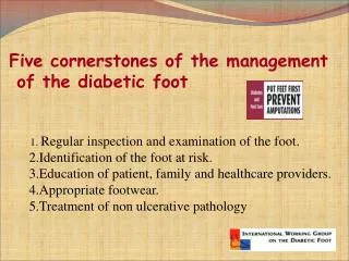
Definition of a Diabetic Foot infection Epidemiology Pathogenesis of a Diabetic Foot Infection classification Assessment Microbiology Principle of antibiotic treatment
Definition of a Diabetic Foot Infection(1) • No generally-accepted definition • Foot infections in diabetics can be ulcer- or non-ulcer related • Anatomic location of primary site • Depth of infection (skin/soft tissue vs. bone/joint) • Isolation of pathogenic bacteria from an appropriate culture specimen
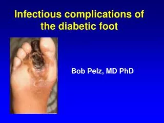
Definition of a Diabetic Foot Infection(2) • entrance ,growth ,metabolic activity and ensuing pathophysiologic effects of microorganisms in the tissues of a patient • Purulent discharge from the ulcer • Signs of inflammation around the ulcer • Systemic signs (fever-leukocytosis) • The manifestation of the inflammatory signs depends on intact nervous and vascular system
Epidemiology • life time risk of DM patient : 15% • 14-20% will need amputation • 1 leg is lost every 30 sec. • More than 80% are potentially preventable • Site of foot ulcers: toes: 51%plantar metatarsal head: 28%dorsum of foot: 14%multiple ulcers: 7%
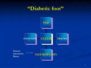
Pathogenesis of diabetic foot infection triangle of devil infection Bad perfusion Badsensation
Classification Systems for Diabetic Foot Infections • Classification systems • Severity of Infection • Foot Ulcer (Wound) • No generally-accepted classification • Differ in criteria & complexity • Require validation for clinical trials
Classification Systems for Severity of Diabetic Foot Infections • Limb-threatening vs. non-limb threatening • Mild, moderate, severe
Classification Systems for Diabetic Foot Ulcers • Wagner • Univ. of Texas • Depth-ischemia class.
Wagner Classification 0- Intact skin (may have bony deformities. 1- Localized superficial ulcer. 2- Deep ulcer to tendon, bone, ligament or joint. 3- Deep abscess or osteomyelitis. 4- Gangrene of toes or forefoot. 5- Gangrene of whole foot. Wagner FW: The diabetic foot and amputations of the foot. In Surgery of the Foot. 5th ed. Mann, R editor. St Louis, Mo. The C.V. Mosby Company.
Depth- ischemia classification Grade 0 no skin change Grade 2exposed tendon, joint Grade A no ischemia Grade Cpartial gangrene Grade 1 superficial ulcer Grade 3 bone exposure Grade Bischemia,no gangrene GradeDcomplete gangrene
Management based classificationstructuredamage • Skin • Subcutaneous tissues • Muscle and tendon • Bone • Articulation Extention of infection Perfusion of the foot • Good • Moderate • Poor • Able to correction or not
Multidisciplinary team • 1-Diabetologist • 2-Vascular surgeon • 3-Orthopedics • 4-Infection disease • 5-Plastic surgeon • 6-Podiatrician
Six intervention demonstrate efficacy in diabetic foot management 1- off loading 2- Debridement and drainage 3- wound dressing 4- appropriate use of antibiotic 5- revascularization 6- limited amputation
Laboratory hematology chemistry HgbA1C C-Reactive Protein Wound, tissue, and blood cultures Wound or ulcer dimensions X ray imaging MRI Isotope scan Doppler Pulse oxygenation measurement (toe) Arteriography Baseline Assessments 1-Extension of infection 2-Vascular assessment 3-General diabetes assess.
Diagnosis of osteomylitis is very important • X Ray is positive after 30-50%of bone destruction(2 weeks) • MRI • CT.Scan • 3-phase bone scan • Leukocyte scan • Guided bone biopsy
Epidemiology Definition of a Diabetic Foot infection Pathogenesis of a Diabetic Foot Infection classification Assessment Microbiology Principle of antibiotic treatment
Microbes and Chronic Wounds • All chronic wounds are contaminated by bacteria. • Wound healing occurs in the presence of bacteria. • It is not the presence of organisms but their interaction with the patient that determines their influence on wound healing.
Louis Pasteur “ The germ is nothing. It is the terrain in which it is found that is everything.” Pasteur, L. (1880) De l’attenuation virus du cholera des poules. CR Acad. Sci. 91: 673-680.
Definitions Wound contamination: the presence of non-replicating organisms in the wound. Wound colonization: the presence of replicating microorganisms adherent to the wound in the absence of injury to the host. Wound Infection: the presence of replicating microorganisms within a wound that cause host injury.
Microbiology of Wounds • The microbial flora in wounds appear to change over time. • Early acute wound; Normal skin flora predominate. • S. aureus, and Beta-hemolytic Streptococcus soon follow. (Group B Streptococcus and S. aureus are common organisms found in diabetic foot ulcers)
Microbiology of Wounds • After about 4 weeks • Facultative anaerobic gram negative rods will colonize the wound. • Most common ones= Proteus, E. coli, and Klebsiella. • As the wound deteriorates deeper structures are affected. Anaerobes become more common. Oftentimes infections are polymicrobial (4-5).
Microbiology of Wounds • In summary: early chronic wounds contain mostly gram-positive organisms. • Wounds of several months duration with deep structure involvement will have on average 4-5 microbial pathogens, including anaerobes (see more gram-negative organisms).
How do you know when a wound is infected? • This can be very difficult. • A continuum exists between when pathogens colonize the wound and then start to cause damage. • There is no absolutely foolproof laboratory test that will aid in this diagnosis.
How do you know when a wound is infected? • One feature is common to all infected chronic wounds; • The failure of the wound to heal and progressive deterioration of the wound. • Unfortunately, wound infections are not the only reasons for poor wound healing.
How do you know when an ulcer is infected? • The typical features of wound infections: • increased exudate • increased swelling • increased erythema • increased pain • increased local temperature • Periwound cellulitis, ascending infection, change in appearance of granulation tissue (discoloration, prone to bleed, highly friable).
Methicillin – resistant Staph. Au. An increasing problem • Retrospective analysis of 63 swabs from infected foot ulcer • Gram+ aerobic 84.2% staph. Au.79% • 30.2% MRSA • Not related to prior antibiotic usage ( dang and al. diab.med.20;2:159 feb2003) • In a prior study MRSA is associated with previous antibiotic treatment (tentolouris and al. diab.med.16;9:767sep1999)
141 microbes isolated from 93 diabetic foot ulcer Study done on syrian population presented in SDA sept2003 B.hammad MD and H.Jammal MD
Epidemiology Definition of a Diabetic Foot infection Pathogenesis of a Diabetic Foot Infection classification Assessment Microbiology Principle of antibiotic treatment
Treatment • Management of infection: 1- antibiotics. 2-Incision and drainage.3-soft tissue, joint and bone resection 4-amputation
What is the best approach? • 1-Oral antibiotic follow up after one week • 2-IV antibiotic in the hospital and observation • 3-Rapid drainage + IVantibiotic
Should we clean uncomplicated foot ulcer with antibiotics? • 44 Clinically uninfected neuropathic foot ulcer • Randomized to amoxi+clav vs. placebo • 20 days follow-up no difference in outcome (chantelau and al. diab. Med. 1996 ;13:156-159) • 64 new foot ulcer with no clinical evidence of infection • Randomized to antibiotics vs. placebo • Patients with ischemia and positive ulcer swabs should be considered for early antibiotic treatment ( foster and al. diab. Med.1998;15:suppl.2)
Principles of treatment • Evidence-based regimes • empirical therapy vs specific therapy • Optimal dosage • Optimal duration • Identification and removal of infective focus • Recognition of adverse effects
The -lactams • Penicillins • penicillin V/G, ampicillin, amoxycillin, cloxacillin, ticarcillin, piperacillin • Cephalosporins • 1st generation e.g. cefazolin, cefalexin (Keflex) • 2nd generation e.g. cefuroxime (Zinacef, Zinnat )
The -lactams • 3rd generation e.g. ceftriaxone (Rocephin ), cefotaxime (Claforan ), ceftazidime (Fortum ), cefoperozone (Cefobid ), ceftibuten (Cedax ) • 4th generation e.g. cefepime (Maxipime ) • Carbapenems • imipenem, meropenem • Monobactam • aztreonam
Macrolides and Quinolones • Macrolides • erythromycin, clarithromycin (Klacid ), azithromycin (Zithromax ) • Quinolones (FQ) • ofloxacin, levofloxacin (Cravit ), Ciprofloxacin (Ciproxin )
Others • Aminoglycosides • gentamicin, amikacin, netromycin* (NA) • Tetracyclines • doxycyline (Vibramycin ), minocycline • Glycopeptides • vancomycin, teicoplanin • New: linezolid, ertapenem, moxifloxacin
Badperfusion ischemic Normal perfusion Non-ischemic deep superficial swab signs of infection No signs of infection swab Largecoverage No antibiotics Gram+ Large coverage
Recent and superficial ulcer or cellulitis (non ischemic) Staph. Au. + strep • Cloxacillin • Amoxi+ with -lactamase inhibitors • Cefazolin • Cephalexin • Clindamycin
Deep ulcer or neuroischemic ulcer polymicrobial: gram positive cocci, gram negative bacilli and anaerobes • -lactam + -lactamase inhibitors +amikacin • 3rd GC + clindamycin • ciprofloxacin + clindamycin • Ciprofloxacin + linezolid • carbapenems vancomycin if life threatening
most ulcers will heal with the traditional Therapy • For low grade uninfected wounds a form of removable or irremovable offloading device should be a part of any treatment plan. The TCC is the most established; • We can not recommend any one dressing over another; • Debridement should still be done the old fashioned way but could be facilitated by using Hydrogel or MDT where available; • if wounds fail to heal, treating them with a skin graft or adding becaplermin (or the platelet releasate) not been validated as cost effective in any clinical trial. • The use of systemic HBO or Iloprost, especially in high grade ulcers with a significant ischaemic element