Download
1 / 26
270 likes | 436 Views
Nordic-Baltic Bifurcation Study III Randomized Comparison of Final Kissing Balloon Dilatation vs. no Final Kissing Balloon Dilatation in Patients with Coronary Bifurcation Lesions Treated With Main Vessel stenting.

E N D
Nordic-Baltic Bifurcation Study IIIRandomized Comparison of Final Kissing Balloon Dilatation vs. no Final Kissing Balloon Dilatation in Patients with Coronary Bifurcation Lesions Treated With Main Vessel stenting MattiNiemela, Kari Kervinen, AndrejsErglis,Niels R. Holm, Michael Maeng, Evald H Christiansen, IndulisKumsars, Sandra Jegere, AndisDombrovskis, PålGunnes, SindreStavnes,TerjeSteigen,ThorTrovik, SailaVikman,MarkkuEskola, HannuRomppanen,TimoMakikallio, Knud N Hansen, Per Thayssen, Lars Åberge,Lisette Jensen, Anders Hervold, J Airaksinen, MikkoPietila, Ole Frobert, Thomas Kellerth, Jan Ravkilde,Jens Aarøe,SteffenHelqvist, IwarSjögren, Stefan James,HeikkiMiettinen, Jens F Lassen, Leif Thuesen For the Nordic-Baltic PCI Study Group
Nordic-Baltic Bifurcation Study IIIParticipating Centers Denmark AarhusUniversityHospital Odense UniversityHospital AalborgUniversityHospital RigshospitaletCopenhagen Latvia Paul StradinsHospital, Riga Sweden Örebro Hospital Uppsala UniversityHospital Falun Hospital Finland Oulu UniversityHospital Tampere UniversityHospital Turku UniversityHospital Kajaani Central Hospital Rovaniemi Central Hospital Kemi Central Hospital Kuopio UniversityHospital Norway FeiringHeartClinic Tromsø University Hospital Rikshopsitalet
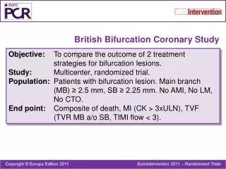
Background The provisional SB stenting strategy has emerged the preferred bifurcation treatment strategy Whether routine Final Kissing Balloon Dilatation (FKBD) after MV stenting improves clinical and angiographic outcome is less well known
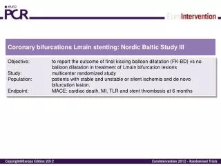
Purpose of the NORDIC III study In a randomized multicenter setting in coronary bifurcations treated with MV stenting using SES to compare No FKBD FKBD • To assess the effect of these strategies to patient outcomes • and angiographic results
Estimate of eligible patients (n= 2385) Randomized patients (n= 477) No FKBD (n= 239) FKBD (n= 238) 6 month clinical FU (n= 239, 100%) 6 month clinical FU (n= 238, 100%) Stratification at randomization Scheduled angiographic FU after 8 months (n= 189) Scheduled angiographic FU after 8 months (n=185) Angiographic FU available (n= 162, 86%) Angiographic FU available (n=164, 88%)
Inclusion criteria Indication Stable angina pectoris Unstable angina pectoris Silent ischaemia Lesion location LAD/diagonal CX/obtuse marginal RCA PDA/postero-lateral branch LM/LAD/CX Vesselsize main vessel diameter ≥ 2.50mm side branch diameter ≥ 2.25 mm
Exclusion criteria ST-segment elevation MI within 24 hours Expected survival< 1 year S-creatinine> 200 µmol/L Allergy to aspirin, clopidogrel, or to sirolimus
The main treatment principles • Wiring of both MV and SB • Predilatation of MV/SB at discretion of the operator • Stenting MV, jailing SB wire • If TIMI flow 3 in MV and SB → Randomization • No-FKBD group: procedure terminated • FKBD group: • 4. rewiring of jailed SB • 5. FKBD • - if SB TIMI flow<3 → SB stenting
- Age, mean+SD 64+10 65+10 ns Male (%) 72.4 73.1 ns Diabetes (%) 16 18 ns Smoking (%) 23 20 ns Hypertension (%) 66 61 ns Statin Tx (%) 84 83 ns Family history (%) 61 56 ns History of PCI (%) 31 24 ns History of CABG (%) 2 3 ns Baseline demographics p value No FKBD FKBD n=239 n=238 +kiss Kissing P value n=238
Coronary angiographyVisual assessment p value No FKBD FKBD Crush Culotte P-value (n=210) (n=215) MV lesion length (mm) 17.7 + 10.2 17.3 + 8.6 ns MV stent length (mm) 22.9 + 10.5 23.6 + 11.1 ns SB lesion length (mm) 3.6 + 4.2 3.4 + 3.9 ns Prx. MV ref. diam. (mm) 3.4 + 0.4 3.4 + 0.6 ns Dis. MV ref. diam. (mm) 3.2 + 0.3 3.2 + 0.4 ns SB ref. diam. (mm) 2.7 + 0.4 2.6 + 0.3 ns n=239 n=238
Reference diameter before procedure on QCA ns mm ns ns
Crush Culotte P-value (n=210) (n=215) SB predilatation, (%) 27.6 29.0 0.76 Final Kissing (%) 0.8 97.1 0.0001 SB dilatation thr. MV 1.7 97.1 0.0001 stent or FKBD SB stented, n (%) 0(0) 3 (1.3) 0.12 Tr. successful*, n (%) 236( 98,7) 236 (99.2) ns Procedure time (min) 47 + 22 61 + 28 0.0001 Fluorosc. time (min) 11 + 10 16 + 12 0.0001 Contrast (ml) 200 + 92 235 + 97 0.0001 Procedure data P value Culotte No FKBD FKBD p-value n=210 n=210 n=238 n=239 *residual stenosis <30% of MV+TIMI III flow in SB
Primary composite end point of MACE (cardiac death, index lesion MI, TLR, stent thrombosis) after 6 months % 2.5 2.1 ns FKBD No - FKBD
CCS angina class > 2 before treatment and at 6-month follow up ns
Quantitative Coronary Analysis (QCA) at 8 month after PCI procedure
Quantitative Coronary Analysis (QCA) Joint angiographic core lab: • -Aarhus University Hospital, Skejby, Denmark • - Paul StradinsClinical Hospital, Riga, Latvia Analysed by computer-based software dedicated to bifurcation analysis (QAngio XA version 7.2, Medis, Leiden, The Netherlands) At 8 months
The patient demographics showed no significant differences between the treatment groups Lesion characteristics
Crush Culotte P-value (n=210) (n=215) SB predilated (%) 31.3 35 ns FKBD (%) 0.8 97.1 0.0001 SB stented (%) 0.0 0.6 ns Tr. successful* (%) 98.8 99.4 ns Procedure data P value Culotte No FKBD FKBD p-value n=210 n=210 n=164 n=162 *residual stenosis <30% of MV+TIMI III flow in SB
(Re)stenosis at 8-months QCA: Entire bifurcation lesion % p=0.11 17.3% 11.0% Binary Restenosis: ≥50% diameter stenosis at follow-up
Restenosis: In-Segment Main Vessel % P=0.68 3.1% 2.5% Binary Restenosis: ≥50% diameter stenosis at follow-up
(Re)stenosis: Ostial Side Branch % p=0.039 15.4% 7.9% Binary Restenosis: ≥50% diameter stenosis at follow-up
Late Lumen Loss mm p=0.42 p=0.23 p=0.34 P=0.42 P=0.23 P=0.52
True bifurcation subgroup at 8 month angio FU Medina classification 1,1,1 - 1,0,1 - 0,1,1 No FKBD FKBD p value In-segment MV ≥50% DS, n (%) 2 (2.2) 3 (3.8) ns SB ≥50% DS, n (%) 16 (20) 7 (7.6) 0.024 n=80 (46.5%) n=92 (53.5%)
True bifurcation subgroupMACE and TLR at 6 month clinical FU % P=0.68 2.5% P=0.62 1.7% 1.7% (n=121) (n=118) 0.8%
Conclusion In coronary bifurcation lesions MV stenting with and without FKBD was associated with similar 6-month clinical outcome The simple no-FKBD strategy was associated to shorter procedure and fluoroscopy time and reduced use of contrast media FKBD reduced angiographic SB (re)stenosis especially in patients with true bifurcation lesion, which was not, however, translated into the clinical outcome