Download

1 / 104
1.13k likes | 1.24k Views
Gastroesophageal Reflux Disease. GERD. By Ahmed Abudeif Abd Elaal Resident in tropical medicine & gastroenterology department. Definitions: Gastroesophageal Reflux (GER): Escape of gastric contents into the esophagus. This process may or may not produce symptoms.

E N D
Gastroesophageal Reflux Disease GERD By Ahmed Abudeif Abd Elaal Resident in tropical medicine & gastroenterology department
Definitions: Gastroesophageal Reflux (GER): Escape of gastric contents into the esophagus. This process may or may not produce symptoms. Reflux esophagitis: Esophageal inflammation caused by the refluxed material. Gastroesophageal Reflux Disease (GERD):Any symptomatic condition or anatomic alteration caused by the reflux of noxious material from the stomach into the esophagus.
Pathogenesis of GERD: • Antireflux mechanisms. B) Gastric Factors. C) Esophageal Clearance Mechanisms. D) Esophageal Epithelial Resistance.
Antireflux Mechanisms: Normally, there is a positive pressure gradient between the abdomen and the thorax that tends to promote the reflux of material from the stomach into the esophagus. In the absence of effective antireflux mechanisms this pressure differential would result in continuous gastroesophageal reflux.
Lower Esophageal Sphincter (LES) It is a 1-3.5 cm segment of specialized circular muscle in the wall of distal esophagus. It maintains a resting pressure of 10-45 mmHg higher than that of the stomach.
Types of LES dysfunction: 1) Intrinsic weakness of the LES muscle: The resting pressure in the LES remains at or near 0. Responsible for > 25% of reflux episodes in patients with severe GERD. 2) Inadequate LES response to increased abdominal pressure.
3) Transient LES relaxation (TLESR): Normally, the LES relaxes for 3-10 seconds to allow the swallowed bolus to enter the stomach. TLESR is not preceded by swallowing & lasts for up to 45 seconds. It is responsible for 70% of reflux episodes in patients with severe GERD. Incompetent LES
2) Crural Diaphragm When the crural diaphragm contract e.g. inspiration the crurae come together & pinch the distal esophagus.
3) Anatomic Features: • The oblique angle of insertion of the esophagus into the stomach (angle of His).
b) The circular muscle fibers of the fundus encircle the lower end of the esophagus. c) The presence of an intraabdominal segment of the esophagus. d) The mucosal rosette of the upper end of the stomach forming a plug to the lower end of the esophagus.
B) Gastric Factors: • Irritant potency of the refluxed material Esophageal injury occurs when the refluxed material is caustic to the esophageal mucosa. Caustic agents that can be found in the stomach include acid, pepsin, bile & pancreatic enzymes.
2) Delayed gastric emptying It causes gastric distention that can stimulate gastric acid secretion & trigger TLESR. Causes: 1- Pyloric channel or duodenal ulcers. 2- Mechanical obstruction e.g. by tumour. 3- Neuromuscular abnormalities e.g. DM, collagen diseases, hypothyroidism, …
C) Esophageal Clearance Mechanisms: The esophagus is cleared of acid by 4 mechanisms: • Gravity. • Peristalsis. 3) Salivation. 4) Intrinsic esophageal bicarbonate production.
Impaired esophageal clearance can occur in: Sleep: due to 1) Elimination of the effect of gravity. 2) Salivation & swallowing cease during sleep “no peristalsis due to absent swallowing”. Scleroderma: due to impaired peristalsis. Cigarette smoking: due to decreased salivation. Hiatus hernia.
D) Esophageal Epithelial Resistance: a) Pre-epithelial defenses: 1) Mucous layer: Acts as a lubricant & a protective barrier against noxious &irritant luminal contents. 2) Unstirred water layer: Lies under the mucous layer & its rich in bicarbonate. It provides a protective alkaline microenvironment. b) Post-epithelial defenses.
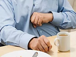
A) Clinical Presentation 1) Heartburn (Pyrosis): Substernal burning sensation radiating to the chest sometimes to the throat or the back. The pain is usually relieved by ingestion of antacids. within 5 minutes The pain is aggravated by - Ingestion of foods that decrease the LES pressure as chocolate, onions, peppermint, coffee, tea & foods that have a high content of fat & sugar.
- Ingestion of foods that irritate the esophageal mucosa directly as spicy foods, citrus products and tomato products. - Practices that increase the intraabdominal pressure e.g. bend over, lift a heavy object, strain to defecate or run.
2) Regurgitation: Reflux of sour or bitter material into the mouth usually at night, when lying down or bending over. It suggests severe reflux.
3) Dysphagia: Difficulty in swallowing. Usually indicates narrowing or stricture of the esophagus. It may occur due to inflammation & oedema.
4) Odynophagia: Pain on swallowing. It suggests the presence of esophageal ulceration.
5) Water brash: Filling of the mouth suddenly with saliva. Its due to reflex salivary secretion stimulated by acid in the esophagus.
6) Chest pain: Resembles anginal pain. This pain may result from: • Acid induced irritation of the nerve endings. • GER-induced esophageal spasm. • GER-induced angina pectoris.
8) Pulmonary symptoms: e.g. chronic cough, hoarseness of voice, wheezing, haemoptysis, asthma & recurrent aspiration pneumonia. The above manifestations may be due to aspiration or vagus mediated neural reflexes.
9) Nighttime GERmay cause: Sleep apnoea. Poor sleep. Insomnia. Daytime sleepiness.
B) Diagnostic tests: Indications: 1) Patients with atypical signs or symptoms. 2) Patients with typical signs & symptoms that don’t respond well to acid suppression.
Endoscopic examination: EGD identifies the presence and severity of esophagitis and the possible presence of Barrett’s esophagus More sensitive than radiology for diagnosis of esophagitis & biopsy can be taken from any abnormal areas. Endoscopic evidence of esophagitis is present in 50-70% of patients with typical history of GERD so a normal EGD (NERD) doesn’t exclude GERD.
The Los Angeles Classification System for the endoscopic assessment of grade of esophagitis
The Los Angeles Classification System for the endoscopic assessment of grade of esophagitis
The Los Angeles Classification System for the endoscopic assessment of grade of esophagitis GRADE A: One or more mucosal breaks no longer than 5 mm, non of which extends between the tops of the mucosal folds
The Los Angeles Classification System for the endoscopic assessment of grade of esophagitis GRADE B: One or more mucosal breaks more than 5 mm long, none of which extends between the tops of two mucosal folds
The Los Angeles Classification System for the endoscopic assessment of grade of esophagitis GRADE C: Mucosal breaks that extend between the tops of two or more mucosal folds, but which involve less than 75% of the oesophageal circumference
The Los Angeles Classification System for the endoscopic assessment of grade of esophagitis GRADE D: Mucosal breaks which involve at least 75% of the oesophageal circumference
Savary-Miller Classification of esophagitis Grade I: Single or multiple erosions are found on a single fold; erosions may be erythematous or exudative.
Savary-Miller Classification of esophagitis Grade II: Multiple erosions affect multiple folds.
Savary-Miller Classification of esophagitis Grade III: Multiple circumferential erosions.
Savary-Miller Classification of esophagitis Grade IV: Ulcer, stricture, and esophageal shortening.
Savary-Miller Classification of esophagitis Grade V:Barrett’s epithelium.
2) Barium swallow: Can reveal: • Signs of esophagitis including: 1- Thickening of the esophageal folds. 2- Erosions. 3- Ulcerations. 4- Strictures. b) GER of barium.
Disadvantages: 1) Less sensitive than endoscopy for demonstrating esophagitis. 2) Biopsy specimens can't be taken.
3) Ambulatory monitoring of esophageal pH: Its used to document the pattern, frequency & duration of acid reflux & to seek correlation between reflux episodes & symptoms. Normally, esophageal pH remains below 4 for less than 4.5% of the 24-hour monitoring period. Acid reflux episode: a drop in esophageal pH below 4 & the total Reflux episodes exceed 5% of the total monitoring time.
4) Acid perfusion (Bernstein) test: It has been used to support acid reflux as the cause of symptoms. The esophagus is perfused with 0.1 N HCl. Reproduction of chest pain with acid perfusion implicates GERD as a cause of chest pain.
5) Histology: Histological changes of esophagitis: 1) Lengthening of the papillae so that they occupy more than 2/3 of the thickness of the squamous mucosa. 2) Hyperplasia of cells in the basal zone so that it occupies more than 15% of the mucosal thickness. 3) Infiltration of the epithelium with eosinophils & PMNLs.
Medical Treatment: a) Lifestyle Modifications. b) Pharmacologic Therapy: 1) H2 blockers 2) Proton Pump Inhibitors. 3) Antacids. 4) Prokinetic Drugs. 5) Sucralfate. B) Antireflux Surgery. C) Endoscopic Antireflux Procedures.
Medical Treatment: • Lifestyle Modifications: 1) Elevation of the head of the bed by 4-6 inch blocks or 6 inch foam-rubber wedge in place of or under the pillow. 2) Weight loss for obese patients. - Obesity increase the intraabdominal pressure.
3) Avoid: a) Smoking - It decrease LES pressure & salivation. b) Alcohol - It decrease LES pressure. c) Fatty meal, chocolate, onion, tea, coffee & carminatives. - They decrease LES pressure & delay gastric emptying.
d) Spicy foods, citrus & tomato products. - They cause direct irritation of the esophageal mucosa. e) Drugs that decrease LES pressure & delay gastric emptying as Ca++ channel blockers, nitrates, drugs that have anticholinergic effects “e.g. phenothiazines, TCA”, theophylline preparations, progesterone & benzodiazepines.