Download
1 / 64
730 likes | 1.34k Views
The Spinal Cord, Nerves and Reflexes. March 24 th , 2008 Martini Chapter 13. Gross Anatomy of the Spinal Cord. adult spinal cord is 18 inches long and 0.55 inches wide cord ends between L1 and L2. Gross Anatomy of the Spinal Cord. posterior median sulcus anterior median sulcus.

E N D
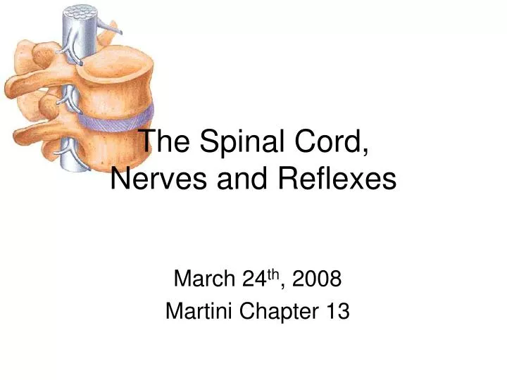
The Spinal Cord, Nerves and Reflexes March 24th, 2008 Martini Chapter 13
Gross Anatomy of the Spinal Cord • adult spinal cord is 18 inches long and 0.55 inches wide • cord ends between L1 and L2
Gross Anatomy of the Spinal Cord • posterior median sulcus • anterior median sulcus
Gross Anatomy of the Spinal Cord • enlargements: • areas of gray matter dedicated to the sensory and motor control of the limbs
Gross Anatomy of the Spinal Cord • cervical enlargement: • supplies nerves to the shoulder and upper limbs
Gross Anatomy of the Spinal Cord • lumbar enlargement: • supplies nerves to the pelvis and lower limbs
Gross Anatomy of the Spinal Cord • conusmedullaris: • inferior to the lumbar enlargement • spinal cord becomes tapered • filumterminale • slender strand of fibrous tissue • from tip of conusmedullaris to sacral vertebrae
Spinal Cord Segments • 31 total • named like vertebrae • division based on origins of spinal nerves
Exiting the Spinal Cord • Dorsal root ganglion • ganglion (cluster of cell bodies) • 1 per segment • contains cell bodies of sensory neurons • dorsal roots: afferent axons • ventral roots: efferent axons • roots pass through the vertebral column at the intervertebral foramen (between vertebrae) • Spinal Nerves: • dorsal and ventral roots combine as they leave vertebral column • classified as “mixed” nerves (i.e., both efferent and afferent fibers)
Naming Spinal Nerves • 31 total • Cervical Nerves • C1 passes btwn skull and atlas • remaining cervical nerves named for inferior vertebrae • so C2 exits between C1 and C2 • although there are only 7 cervical vertebrae, there are 8 cervical spinal nerves
Naming Spinal Nerves • 31 total • Thoracic Nerves and below • spinal nerve takes name from superior vertebrae • so T1 spinal nerve exits between T1 and T2
Naming Spinal Nerves • 31 total • Below L1 and L2 • dorsal and ventral roots of segments L2 to S5 must travel inferiorly, past the conusmedullaris to exit the vertebrae. • The combination of these roots and the filumterminale looks like a horses tail – hence the name caudaequina
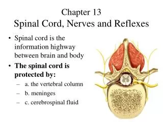
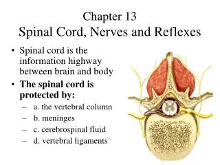
Spinal Meninges • continuous with cranial meninges surrounding the brain • 3 layers • dura mater • arachnoid mater • pia mater • function to provide shock absorption and physical stability
Spinal Meninges: Dura Mater • literally “hard mother” • dense collagen fibers parallel to cord • epidural space • between dura mater and walls of vertebral canal • contains areolar tissue, blood vessels and adipose tissue
Spinal Meninges: Dura Mater • spinal dura mater is only attached at each side of vertebral canal, whereas cranial dura mater fuses with periosteum of occipital bone
Spinal Meninges: Dura Mater • Longitudinal stability: • cranial dura mater fuses with periosteum of occipital bone • within the sacral canal, spinal dura mater becomes dense cord of collagen fibers blending with filumterminale to make the coccygeal ligament • coccygeal ligament ultimately blends with periosteum of coccyx
Spinal Meninges: Dura Mater • Lateral stability: • areolar tissue + adipose tissue in the epidural space • dura mater extends into intervertebral foramen and fuses with connective tissue surrounding nerves
Local Anesthesia • peripheral application of anesthetic • applied locally (at site of injury, or surgical manipulation)
Spinal Anesthesia • central application • applied around spinal cord (usually epidurally) • effects only spinal nerves in the immediate area • mainly sensory anesthesia • difficult in C and T area due to little space • lower lumbar or sacral injections used during childbirth
Spinal Meninges: Arachnoid Mater • Literally “Spider Mother” • arachnoid membrane • simple squamous epithelia line the surface between the dura mater and arachnoid mater • arachnoidtrabeculae • network of collagen and elastic fibers extends between membrane and outer surface of pia mater • subarachnoid space • between arachnoid mater and pia mater, filled with cerebrospinal fluid
Spinal Meninges: Arachnoid Mater • extends as far as the filum terminale • dorsal/ventral roots of cauda equine lie within the subarachnoid space
Spinal Tap • CSF is taken from the subarachnoid space of the inferior lumbar region
Spinal Meninges: Pia Mater • Literally “Delicate Mother” • meshwork of elastic and collagen fibers firmly bound to neural tissue • interwoven with fibers in subarachnoid space • blood vessels that serve spine run along the surface of the pia matter within the subarachnoid space
Stabilizing the Spinal Cord • prevention of side-to-side movement • denticulate ligaments • extend from pia mater through the arachnoid mater to the dura mater • prevention of up/down movement • dural connections • at foramen magnum/occipital bone & at coccygeal ligament
Sectional Anatomy • anterior median fissure & posterior median sulcus mark the left/right division • White matter (Axons, wrapped in fatty myelin look white) • Gray matter (Cell Bodies, less fat, don’t look as white) • horns project toward outer surface • Central Canal • H or butterfly shape
Organization of Gray Matter • Nuclei • groups of specialized neurons • don’t confuse this term with the plural of the nucleus of the cell! • Sensory Nuclei • receive input from periphery • Motor Nuclei • issue motor commands to periphery • Nuclei arranged topographically medial lateral (e.g., medial serves shoulder, lateral serves fingers)
Organization of Gray Matter • Sensory Nuclei • Posterior location • posterior gray horns • somatic sensory nuclei • visceral sensory nuclei
Organization of Gray Matter • Motor Nuclei • Anterior location • anterior gray horns • somatic motor nuclei • Mid-lateral location • lateral gray horn • visceral motor nuclei • only in T and L
Organization of Gray Matter • Gray Commissures (joining together) • posterior and anterior to central canal • contain axons that cross the spinal cord
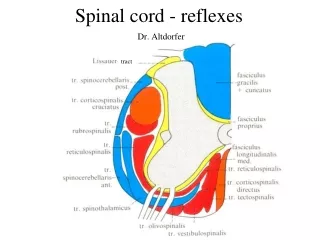
Organization of White Matter • Arranged in Columns (funiculi) • posterior white column • btwn post. gray horn and sulcus • anterior white column • btwn ant. gray horn and fissure • interconnected by anterior white commissure • lateral white column • btwn ant. and post. columns
Organization of White Matter • What are the Columns? • tracts or fasciculus (bundles) of axons • carry similar information (sensory or motor) • all go in the same direction (up or down) • Ascending tracts • spinal cord brain • sensory • Descending tracts • brain spinal cord • motor
Organization of White Matter • All tracts are paired • Most cross over (decussate) at some point • Most consist of a chain of 2 or 3 successive neurons
White Matter Tracts • Named for their origin and destination • We will learn more about these in Chps 15-16
Spinal Nerves • Surrounded by Connective Tissue • Epineurium • dense collagen network • arteries and veins enter here • Perineurium • divide nerve into fascicles (bundles of axons) • arteries and veins branch here • Endoneurium • surrounds each individual axon • capillaries supply axons • These tissues extend and layer the peripheral nerves as well.
Peripheral Distribution of Spinal Nerves • Each spinal nerve connects to the spinal cord through to roots. • Ventral roots arise from the anterior horn and contain motor (efferent) fibers • Dorsal roots arise from sensory neurons in the dorsal root ganglion and contain sensory (afferent) fibers
Peripheral Distribution of Spinal Nerves • Dorsal Root Ganglion: • contains cell bodies of afferent sensory neurons • The dorsal and ventral roots join to form the spinal nerve, which then branches into rami.
Rami of the Ventral (Motor) Root • RamiCommunicantes • visceral motor output via sympathetic ganglion and becoming sympathetic nerve • white ramus (myelin) • gray ramus (no myelin) • Dorsal Rami • visceral and somatic motor input to skin and back • Ventral Rami (larger) • visceral and somatic motor input to structures in body wall and limbs
Rami of the Dorsal (Sensory) Root • Dorsal Rami • sensory (somatic and visceral) input from back • White Rami • sensory (visceral only) input from organs • Ventral Rami • sensory input (somatic and visceral) from structures in body wall and limbs
Nerve Plexus • All ventral rami (except T2-12) form complex networks of nerves called plexuses (braid) Four Major Plexuses • Cervical Plexus • Brachial Plexus • Lumbar Plexus • Sacral Plexus
The Cervical Plexus • ventral rami of C1-5 • Most branches are cutaneous nerves of the neck, ear, back of head, and shoulders • Phrenic nerve • major motor and sensory nerve of the diaphragm
The Brachial Plexus • Formed by the ventral rami of C5-C8 and T1 • C4 and T2 may also contribute • It gives rise to the nerves that innervate the upper limb
Trunks and Cords of Brachial Plexus Nerves that form brachial plexus originate from: Trunks - large bundles of axons from several spinal nerves superior, middle, and inferior Cords - smaller branches that originate at trunks lateral, medial, and posterior cords
Specific Nerves of the Brachial Plexus • ulnar nerve (medial cord) • supplies the flexor carpiulnaris and part of the flexor digitorumprofundus • radial nerve (posterior cord) • innervates essentially all extensor muscles
The Lumbar Plexus Formed by the ventral rami of T12-L4 innervates the thigh, abdominal wall, and psoas muscle Femoral Nerve innervates anterior thigh, hip flexors/adductors, skin over anterior thigh andmedial leg/foot
The Lumbar Plexus Formed by the ventral rami of T12-L4 innervates the thigh, abdominal wall, and psoas muscle Femoral Nerve innervates anterior thigh, hip flexors/adductors, skin over anterior thigh andmedial leg/foot
The Sacral Plexus Formed by the ventral rami of L4-S4 innervates the buttock, lower limb, pelvic structures, and the perineum sciatic nerve the longest and thickest nerve of the body actually two nerves tibial nerve common fibular nerve
The Sacral Plexus Formed by the ventral rami of L4-S4 innervates the buttock, lower limb, pelvic structures, and the perineum sciatic nerve the longest and thickest nerve of the body actually two nerves tibial nerve common fibular nerve
Dermatome • Area of skin innervated by the cutaneous branches of a single spinal nerve. • All segments except C1 have dermotomal distribution • upper extremities: C5-T1 • lower extremities: L1-S1 • Clinical Significance: • loss of sensation can indicate specific damage/infection of spinal nerve or DRG
Neural Circuits:information processing Divergence: spreads stimulation to many neurons or neuronal pools in CNS typical of sensory input to CNS Figure 13–13a
Neural Circuits:information processing Convergence: brings input from many sources to single neuron typical of control of motor output conscious and subconscious control of muscle contraction converge onto the same motor neuron Figure 13–13a