Download
1 / 22
410 likes | 1k Views
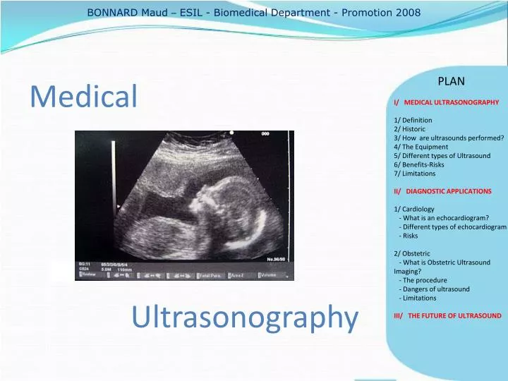
BONNARD Maud – ESIL - Biomedical Department - Promotion 2008. PLAN. Medical Ultrasonography. I/ MEDICAL ULTRASONOGRAPHY 1/ Definition 2/ Historic 3/ How are ultrasounds performed ? 4/ The Equipment 5/ Different types of Ultrasound 6/ Benefits-Risks 7/ Limitations

E N D
BONNARD Maud – ESIL - Biomedical Department - Promotion 2008 PLAN Medical Ultrasonography • I/ MEDICAL ULTRASONOGRAPHY • 1/ Definition • 2/ Historic • 3/ How are ultrasounds performed? • 4/ The Equipment • 5/ Different types of Ultrasound • 6/ Benefits-Risks • 7/ Limitations • II/ DIAGNOSTIC APPLICATIONS • 1/ Cardiology • - Whatis an echocardiogram? • - Different types of echocardiogram • - Risks • 2/ Obstetric • - What is Obstetric Ultrasound Imaging? • - Theprocedure • - Dangers of ultrasound • - Limitations • III/ THE FUTURE OF ULTRASOUND
I/ MEDICAL ULTRASONOGRAPHY • 1/ Definition • 2/ Historic • 3/ How are ultrasounds performed? • 4/ The Equipment • 5/ Different types of Ultrasound • 6/ Benefits-Risks • 7/ Limitations • II/ DIAGNOSTIC APPLICATIONS • 1/ Cardiology • - Whatis an echocardiogram? • - Different types of echocardiogram • - Risks • 2/ Obstetric • - What is Obstetric Ultrasound Imaging? • - Theprocedure • - Dangers of ultrasound • - Limitations • III/ THE FUTURE OF ULTRASOUND
MEDICAL ULTRASONOGRAPHY – Definition PLAN ULTRASOUNDS = highfrequencysoundwaves • I/ MEDICAL • ULTRASONOGRAPHY • 1/ Definition • 2/ Historic • 3/ How are ultrasounds performed? • 4/ The Equipment • 5/ Different types of Ultrasound • 6/ Benefits-Risks • 7/ Limitations • II/ DIAGNOSTIC APPLICATIONS • 1/ Cardiology • - Whatis an echocardiogram? • - Different types of echocardiogram • - Risks • 2/ Obstetric • - What is Obstetric Ultrasound Imaging? • - Theprocedure • - Dangers of ultrasound • - Limitations • III/ THE FUTURE OF ULTRASOUND Medicalultrasonography = ultrasound-based diagnostic imaging technique. Muscles and internalorgans ( size, structures, pathologies or lesions). High frequencysoundwaves and theirechoes VISUALIZE
MEDICAL ULTRASONOGRAPHY – Historic PLAN Starts in the mid 19 century by measuring distance under water using sound waves. • I/ MEDICAL • ULTRASONOGRAPHY • 1/ Definition • 2/ Historic • 3/ How are ultrasounds performed? • 4/ The Equipment • 5/ Different types of Ultrasound • 6/ Benefits-Risks • 7/ Limitations • II/ DIAGNOSTIC APPLICATIONS • 1/ Cardiology • - Whatis an echocardiogram? • - Different types of echocardiogram • - Risks • 2/ Obstetric • - What is Obstetric Ultrasound Imaging? • - Theprocedure • - Dangers of ultrasound • - Limitations • III/ THE FUTURE OF ULTRASOUND 1940 Sonar extensively developed and used to detectsubmarines. 1953 First successfulmeasurement of heartactivity . 1964 It became possible to studypregnancyfromstart to finish and diagnose complications. 1975 Invention of 2D and real time ultrasound. 1994 Invention of the 3D ultrasound …
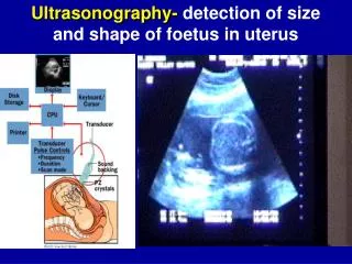
MEDICAL ULTRASONOGRAPHY – How are ultrasoundperformed ? PLAN Gel smeared on the area of the body to undergo the ultrasound • I/ MEDICAL • ULTRASONOGRAPHY • 1/ Definition • 2/ Historic • 3/ How are ultrasound s • performed? • 4/ The Equipment • 5/ Different types of Ultrasound • 6/ Benefits-Risks • 7/ Limitations • II/ DIAGNOSTIC APPLICATIONS • 1/ Cardiology • - Whatis an echocardiogram? • - Different types of echocardiogram • - Risks • 2/ Obstetric • - What is Obstetric Ultrasound Imaging? • - Theprocedure • - Dangers of ultrasound • - Limitations • III/ THE FUTURE OF ULTRASOUND 4. Calculations by a computer 3. Theirechoes are received 2. High frequencysoundwaves sent 5. Dynamic images
MEDICAL ULTRASONOGRAPHY – How are ultrasoundperformed ? PLAN At a given temperature, the sound velocity is constant in a given material, but it varies if the materials are different. • I/ MEDICAL • ULTRASONOGRAPHY • 1/ Definition • 2/ Historic • 3/ How are ultrasound s • performed? • 4/ The Equipment • 5/ Different types of Ultrasound • 6/ Benefits-Risks • 7/ Limitations • II/ DIAGNOSTIC APPLICATIONS • 1/ Cardiology • - Whatis an echocardiogram? • - Different types of echocardiogram • - Risks • 2/ Obstetric • - What is Obstetric Ultrasound Imaging? • - Theprocedure • - Dangers of ultrasound • - Limitations • III/ THE FUTURE OF ULTRASOUND • Like many organs, the tennis ball is solid on the outside and hollow on the inside. • Solid structures (bones, muscles) • reflect sound waves . • show up as white in an • ultrasound image. • Soft or hollow areas (tissues, • chambers of the heart) • don’t reflect sound waves. • appear as black.
MEDICAL ULTRASONOGRAPHY – The Equipment PLAN • I/ MEDICAL • ULTRASONOGRAPHY • 1/ Definition • 2/ Historic • 3/ How are ultrasounds performed? • 4/ The Equipment • 5/ Different types of Ultrasound • 6/ Benefits-Risks • 7/ Limitations • II/ DIAGNOSTIC APPLICATIONS • 1/ Cardiology • - Whatis an echocardiogram? • - Different types of echocardiogram • - Risks • 2/ Obstetric • - What is Obstetric Ultrasound Imaging? • - Theprocedure • - Dangers of ultrasound • - Limitations • III/ THE FUTURE OF ULTRASOUND CPU : central processing unit Console Transducers
MEDICAL ULTRASONOGRAPHY – Different types of ultrasound PLAN • 3D Ultrasound Imaging • I/ MEDICAL • ULTRASONOGRAPHY • 1/ Definition • 2/ Historic • 3/ How are ultrasounds performed? • 4/ The Equipment • 5/ Different types of • Ultrasound • 6/ Benefits-Risks • 7/ Limitations • II/ DIAGNOSTIC APPLICATIONS • 1/ Cardiology • - Whatis an echocardiogram? • - Different types of echocardiogram • - Risks • 2/ Obstetric • - What is Obstetric Ultrasound Imaging? • - Theprocedure • - Dangers of ultrasound • - Limitations • III/ THE FUTURE OF ULTRASOUND 3D ultrasound images • Doppler Ultrasound Power Doppler ultrasound of the kidney.
MEDICAL ULTRASONOGRAPHY – Benefits vsRisks PLAN • Benefits • I/ MEDICAL • ULTRASONOGRAPHY • 1/ Definition • 2/ Historic • 3/ How are ultrasounds performed? • 4/ The Equipment • 5/ Different types of Ultrasound • 6/ Benefits-Risks • 7/ Limitations • II/ DIAGNOSTIC APPLICATIONS • 1/ Cardiology • - Whatis an echocardiogram? • - Different types of echocardiogram • - Risks • 2/ Obstetric • - What is Obstetric Ultrasound Imaging? • - Theprocedure • - Dangers of ultrasound • - Limitations • III/ THE FUTURE OF ULTRASOUND • No ionizing radiation X- RAYS • Non invasive and painless • Easy-to-use • Lessexpensivethanotherimagingmethods • Providesreal-time imaging • Risks • No confirmed adverse biological effects caused by exposures to ultrasound
MEDICAL ULTRASONOGRAPHY – Limitations PLAN • Ultrasoundwavesdo not passthroughair or gas, canbereflected evaluationof the stomach, small intestine and large intestine limited • Ultrasoundhas difficultypenetratingbone onlysee the outer surface of bony structures but not within. • I/ MEDICAL • ULTRASONOGRAPHY • 1/ Definition • 2/ Historic • 3/ How are ultrasounds performed? • 4/ The Equipment • 5/ Different types of Ultrasound • 6/ Benefits-Risks • 7/ Limitations • II/ DIAGNOSTIC APPLICATIONS • 1/ Cardiology • - Whatis an echocardiogram? • - Different types of echocardiogram • - Risks • 2/ Obstetric • - What is Obstetric Ultrasound Imaging? • - Theprocedure • - Dangers of ultrasound • - Limitations • III/ THE FUTURE OF ULTRASOUND
DIAGNOSTIC APPLICATIONS PLAN MANY !!!! • I/ MEDICAL ULTRASONOGRAPHY • 1/ Definition • 2/ Historic • 3/ How are ultrasounds performed? • 4/ The Equipment • 5/ Different types of Ultrasound • 6/ Benefits-Risks • 7/ Limitations • II/ DIAGNOSTIC • APPLICATIONS • 1/ Cardiology • - Whatis an echocardiogram? • - Different types of echocardiogram • - Risks • 2/ Obstetric • - What is Obstetric Ultrasound Imaging? • - Theprocedure • - Dangers of ultrasound • - Limitations • III/ THE FUTURE OF ULTRASOUND • Echocardiogram : heart and its valves, heart's pumping ability • Obstetrical ultrasound : follow development of the fetus • Abdominal ultrasound : detect abnormalities in abdominal organs • (kidneys, liver, pancreas, gallbladder…) • Renal ultrasound : examine the kidneys and urinary tract. • Vascular ultrasound : see the vascular system and its function, detection of blood clots. • …
DIAGNOSTIC APPLICATIONS– Cardiology PLAN Whatis an echocardiogram ? • I/ MEDICAL ULTRASONOGRAPHY • 1/ Definition • 2/ Historic • 3/ How are ultrasounds performed? • 4/ The Equipment • 5/ Different types of Ultrasound • 6/ Benefits-Risks • 7/ Limitations • II/ DIAGNOSTIC APPLICATIONS • 1/ Cardiology • - Whatis an • echocardiogram? • - Different types of echocardiogram • - Risks • 2/ Obstetric • - What is Obstetric Ultrasound Imaging? • - Theprocedure • - Dangers of ultrasound • - Limitations • III/ THE FUTURE OF ULTRASOUND • Ultrasound of the heart • 2D pictures of cardiovascular system • Assessvelocity of blood (doppler ultrasound) • Size, shape of the heart, pumpingstrength. • Anycardiac valve disfonction(leak, regurgitation of blood) • Detectabnormalitieslinked to deseases(coronary artery ) Advantages : noninvasive, no sideeffects
DIAGNOSTIC APPLICATIONS– Cardiology PLAN • Transthoracic echocardiogram (TTE) • Views of the heart obtained by moving the transducer to different • locations on the chest or abdominal wall. • Stress echocardiogram • An echocardiogram is done both before and after the heart is stressed • by having the patient exercise or by injecting a medicine. • To detect coronary artery diseases • Doppler echocardiogram • To look how blood flows through the heart chambers, heart valves, • and blood vessels. • Measures the direction and speed of blood through blood vessels. • Transesophageal echocardiogram (TEE) • Shows clearer pictures of the heart. • (The probe is closer to the heart and the lungs bones of the chest • wall do not block the sound waves) • I/ MEDICAL ULTRASONOGRAPHY • 1/ Definition • 2/ Historic • 3/ How are ultrasounds performed? • 4/ The Equipment • 5/ Different types of Ultrasound • 6/ Benefits-Risks • 7/ Limitations • II/ DIAGNOSTIC APPLICATIONS • 1/ Cardiology • - Whatis an echocardiogram? • - Different types of • echocardiogram • - Risks • 2/ Obstetric • - What is Obstetric Ultrasound Imaging? • - Theprocedure • - Dangers of ultrasound • - Limitations • III/ THE FUTURE OF ULTRASOUND
DIAGNOSTIC APPLICATIONS– Cardiology PLAN • I/ MEDICAL ULTRASONOGRAPHY • 1/ Definition • 2/ Historic • 3/ How are ultrasounds performed? • 4/ The Equipment • 5/ Different types of Ultrasound • 6/ Benefits-Risks • 7/ Limitations • II/ DIAGNOSTIC APPLICATIONS • 1/ Cardiology • - Whatis an echocardiogram? • - Different types of echocardiogram • - Risks • 2/ Obstetric • - What is Obstetric Ultrasound Imaging? • - Theprocedure • - Dangers of ultrasound • - Limitations • III/ THE FUTURE OF ULTRASOUND • No risksfrom a transthoracic or doppler echocardiogram. • Stress echocardiogram : dizziness, lowblood pressure, • shortness of breath, nausea, irregular heartbeats, and heart • attack. • Transesophageal echocardiogram : sometimes nausea, • mouth and throat discomfort, minor bleeding, trouble • breathing, slow or abnormal heartbeats. No risks due to ultrasoundsitselves
DIAGNOSTIC APPLICATIONS– Obstetric PLAN • I/ MEDICAL ULTRASONOGRAPHY • 1/ Definition • 2/ Historic • 3/ How are ultrasounds performed? • 4/ The Equipment • 5/ Different types of Ultrasound • 6/ Benefits-Risks • 7/ Limitations • II/ DIAGNOSTIC APPLICATIONS • 1/ Cardiology • - Whatis an echocardiogram? • - Different types of echocardiogram • - Risks • 2/ Obstetric • - What is Obstetric • Ultrasound Imaging? • - Theprocedure • - Dangers of ultrasound • - Limitations • III/ THE FUTURE OF ULTRASOUND Ultrasound is commonly used to: • Measure the size of the fetus to determine the due date • Determine the position of the fetus • See the number of fetuses in the uterus • Check the fetus' growth rate • See tumors of the ovary and breast • …
DIAGNOSTIC APPLICATIONS– Obstetric PLAN • Information recomposed into a dynamic picture (sonogram) on the monitor screen. • I/ MEDICAL ULTRASONOGRAPHY • 1/ Definition • 2/ Historic • 3/ How are ultrasounds performed? • 4/ The Equipment • 5/ Different types of Ultrasound • 6/ Benefits-Risks • 7/ Limitations • II/ DIAGNOSTIC APPLICATIONS • 1/ Cardiology • - Whatis an echocardiogram? • - Different types of echocardiogram • - Risks • 2/ Obstetric • - What is Obstetric • Ultrasound Imaging? • - Theprocedure • - Dangers of ultrasound • - Limitations • III/ THE FUTURE OF ULTRASOUND • Assess fetal heart beat (doppler ultrasound) and malformations in the fetus. • Make measurements(gestational age, size and growth in the fetus). • Color Doppler clearlydepict the blood flow in fetalbloodvessels in a realtime scan. • (differentcolors = different directions) • Used : • in diagnosis of fetalcardiac and blood • vesselsdefects • in the assessment of the hemodynamic • responses to fetal hypoxia and anemia.
DIAGNOSTIC APPLICATIONS– Obstetric PLAN The procedure is done by : • I/ MEDICAL ULTRASONOGRAPHY • 1/ Definition • 2/ Historic • 3/ How are ultrasounds performed? • 4/ The Equipment • 5/ Different types of Ultrasound • 6/ Benefits-Risks • 7/ Limitations • II/ DIAGNOSTIC APPLICATIONS • 1/ Cardiology • - Whatis an echocardiogram? • - Different types of echocardiogram • - Risks • 2/ Obstetric • - What is Obstetric • Ultrasound Imaging? • - Theprocedure • - Dangers of ultrasound • - Limitations • III/ THE FUTURE OF ULTRASOUND • Placing the transducer (that emits high frequency sound waves) • on the maternal abdomen. • Moving it to "look at" (likened to a light shined from a torch) any • particular content of the uterus. • Repetitive arrays of ultrasound beams scan the fetus in thin slices and are reflected back onto the same transducer.
DIAGNOSTIC APPLICATIONS– Obstetric PLAN Major dangers withultrasound: • I/ MEDICAL ULTRASONOGRAPHY • 1/ Definition • 2/ Historic • 3/ How are ultrasounds performed? • 4/ The Equipment • 5/ Different types of Ultrasound • 6/ Benefits-Risks • 7/ Limitations • II/ DIAGNOSTIC APPLICATIONS • 1/ Cardiology • - Whatis an echocardiogram? • - Different types of echocardiogram • - Risks • 2/ Obstetric • - What is Obstetric • Ultrasound Imaging? • - Theprocedure • - Dangers of ultrasound • - Limitations • III/ THE FUTURE OF ULTRASOUND • Development of heat: • Tissues or water absorb the ultrasound energy • whichincreases their temperature locally. • Formation of bubbles (cavitation): • When dissolved gases come out of solution due to local • heat caused by ultrasound. BUT : No substantiatedill-effects of ultrasounddocumented in studies in eitherhumans or animals.
DIAGNOSTIC APPLICATIONS– Obstetric PLAN Obstetric ultrasound cannot identify all fetal abnormalities • I/ MEDICAL ULTRASONOGRAPHY • 1/ Definition • 2/ Historic • 3/ How are ultrasounds performed? • 4/ The Equipment • 5/ Different types of Ultrasound • 6/ Benefits-Risks • 7/ Limitations • II/ DIAGNOSTIC APPLICATIONS • 1/ Cardiology • - Whatis an echocardiogram? • - Different types of echocardiogram • - Risks • 2/ Obstetric • - What is Obstetric • Ultrasound Imaging? • - Theprocedure • - Dangers of ultrasound • - Limitations • III/ THE FUTURE OF ULTRASOUND If suspicions for a possible abnormality OTHER TESTS ! Amniocentesis (Evaluation of fluid taken from the sac surrounding the baby)
THE FUTURE OF ULTRASOUNDS PLAN Ultrasonography can still be improved ! • I/ MEDICAL ULTRASONOGRAPHY • 1/ Definition • 2/ Historic • 3/ How are ultrasounds performed? • 4/ The Equipment • 5/ Different types of Ultrasound • 6/ Benefits-Risks • 7/ Limitations • II/ DIAGNOSTIC APPLICATIONS • 1/ Cardiology • - Whatis an echocardiogram? • - Different types of echocardiogram • - Risks • 2/ Obstetric • - What is Obstetric • Ultrasound Imaging? • - Theprocedure • - Dangers of ultrasound • - Limitations • III/ THE FUTURE OF • ULTRASOUND • Improve the ultrasound machines technology • Improve the imaging techniques. • Faster, smaller • Betterstoragecapacity • Transducers smaller • more insertable • Develop 3D ultrasound • Developultrasoundimaging + • virtual reality-type displays • help for amniocentesis, biopsy.
CONCLUSION PLAN ULTRASONOGRAPHY : • I/ MEDICAL ULTRASONOGRAPHY • 1/ Definition • 2/ Historic • 3/ How are ultrasounds performed? • 4/ The Equipment • 5/ Different types of Ultrasound • 6/ Benefits-Risks • 7/ Limitations • II/ DIAGNOSTIC APPLICATIONS • 1/ Cardiology • - Whatis an echocardiogram? • - Different types of echocardiogram • - Risks • 2/ Obstetric • - What is Obstetric Ultrasound Imaging? • - Theprocedure • - Dangers of ultrasound • - Limitations • III/ THE FUTURE OF ULTRASOUND • Veryused and usefultechnic for diagnostic • applications in medecine. • Variousforms allow to seemanythings • Benefits and limitations but no riskknown . • Can stillbeimproved to help evenbetterpreventing • deseases or curing people. HOWEVER :
PLAN Thankyou for your attention ! • I/ MEDICAL ULTRASONOGRAPHY • 1/ Definition • 2/ Historic • 3/ How are ultrasounds performed? • 4/ The Equipment • 5/ Different types of Ultrasound • 6/ Benefits-Risks • 7/ Limitations • II/ DIAGNOSTIC APPLICATIONS • 1/ Cardiology • - Whatis an echocardiogram? • - Different types of echocardiogram • - Risks • 2/ Obstetric • - What is Obstetric Ultrasound Imaging? • - Theprocedure • - Dangers of ultrasound • - Limitations • III/ THE FUTURE OF ULTRASOUND Some DJs start learning their craft at an early age...