Download
1 / 59
600 likes | 817 Views
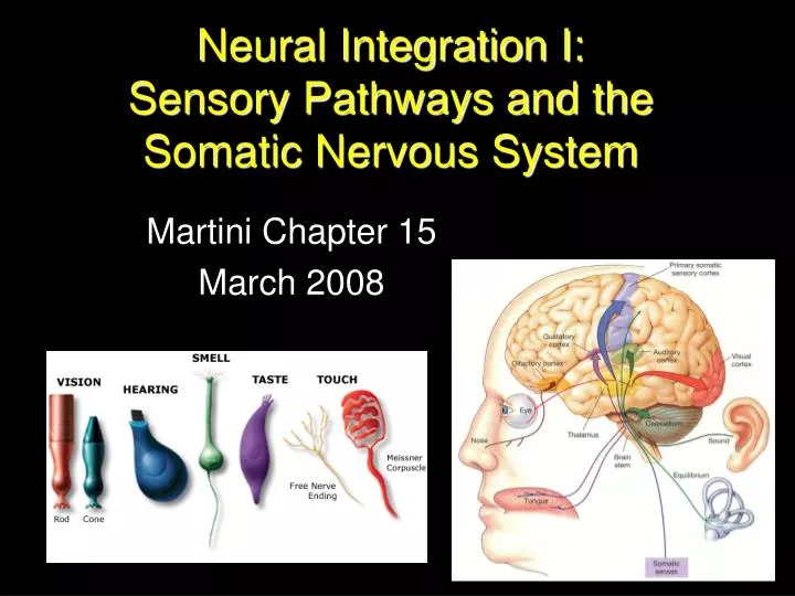
Neural Integration I: Sensory Pathways and the Somatic Nervous System. Martini Chapter 15 March 2008. General Senses. Sensation of temperature, pain, touch, pressure, vibration, and proprioception Sensation : Information provided by sensory receptors to the CNS.

E N D
Neural Integration I: Sensory Pathways and the Somatic Nervous System Martini Chapter 15 March 2008
General Senses • Sensation of temperature, pain, touch, pressure, vibration, and proprioception Sensation: Information provided by sensory receptors to the CNS. Perception: The conscious awareness of sensation.
Special Senses • More complex than general senses, having sensory organs that protect receptors. • olfaction (smell) • vision • gustation (taste) • equilibrium (balance) • hearing
Free Nerve Endings • Simplest type of Sensory receptor • Dendrites of Sensory Neurons detect stimuli • No accessory structures, so they detect numerous types of stimuli (low receptor specificity)
Sensory Receptors • Detects stimuli and translates into action potential that can be conveyed to the CNS in a process called transduction. • Can humans transduce all stimuli?
Transduction • receptor potential • transduction begins when a stimulus changes the transmembrane potential of a sensory neuron • graded effect: stronger stimulus = larger receptor potential. • generator potential • when the receptor potential exceeds the threshold and an action potential is fired
Tonic and Phasic Receptors • Tonic Receptors • always active, indicative of background level of stimulation • change in frequency of action potentials indicates increase or decrease of stimulation • Phasic Receptors • normally inactive • activated for short period by a stimulus • provide information about the intensity and rate of change of stimuli
Specificity of Sensory Receptors • Most receptors detect a specific type of stimulus • due to structure of receptor and presence of other accessory cells surrounding receptor
Receptive Field • The area is monitored by a single receptor cell • The larger the receptive field, the more difficult it is to localize a stimulus
Labeled Lines • The link between the peripheral receptor and the cortical neuron. • Where the labeled line arrives in the cortex determines what you perceive • If the labeled line is activated at a point beyond the receptor it can cause perceptions in the absence of sensation: • seeing halos when you rub your eyes is an example of how our perception of reality is sometimes misleading.
Sensory Adaptation • Reduction in sensitivity of a receptor in the presence of a constant stimulus • Peripheral Adaptation: • level of receptor activity decreases • Central Adaptation: • signal decreases along sensory pathway after receptor • usually inhibition of nuclei along pathway by other neurons
Sensory Adaptation • fast-adapting receptors • phasic receptors • high level of peripheral adaptation • example is thermoreceptors • you are not constantly aware of temperature, only change in temperature
Classification by location exteroreceptors touch, pressure, pain, special senses interoreceptors chemical changes, stretching of tissues, temperature proprioceptors Location of self only in skeletal muscle, tendons, joints, ligaments, & CT coverings of bones & muscles Classification by sensation nociceptors pain thermoreceptors temperature mechanoreceptors deformed by force touch, pressure (BP), vibration, stretch, itch chemoreceptors chemicals in solution Smell, taste, blood chemistry General Senses
Nociceptors • Free nerve endings with large receptive fields • May be sensitive to: • extremes of temperature • mechanical damage • dissolved chemicals, such as chemicals released by injured cells • Very strong stimuli may activate all 3 receptor types.
Nociceptors • Are common in the: • superficial portions of the skin • joint capsules • within the periostea of bones • around the walls of blood vessels • slow-adapting tonic receptors • Two types of Axons carry painful sensations: • Type A fibers • Type C fibers • Carried on pathway to thalamus, reticular formation, primary sensory cortex
Fast Pain • carried by large myelinated Type A fibers • reach CNS quickly and trigger fast reflex • specific activation of primary sensory cortex • easy to localize where pain occurred
Slow Pain • carried by small unmyelinated Type C fibers • reaches CNS slowly • generalized activation of the thalamus • difficult to localize area of pain
The Perception of Pain • chronic pain can lead to facilitation of pain pathways making lesser stimuli seem more painful. • damage to the sensory neuron can lead to the perception of pain in the absence of a painful stimulus
Neurotransmitters and Pain • Sensory Neurons • glutamate and Substance P • excitatory • facilitate neurons along pain pathway
Neurotransmitters and Pain • Natural painkillers • endorphins and enkephalins (in small red neuron) • inhibit the perception of pain by the CNS by inhibiting the release of Substance P from sensory neurons Keep in mind: The perception of pain may be gone, but the stimulus remains.
Thermoreceptors • free nerve endings • dermis • skeletal muscles • liver • hypothalamus • fast-adapting phasic receptors • cold receptors are 3-4X more numerous than warm receptors • carried over same pathways as pain • taken to thalamus, reticular formation and primary sensory cortex
Mechanoreceptors • sensitive to distortions of cell membrane • mechanically gated ion channels open • 3 classes • Tactile receptors • touch, pressure, and vibration • Baroreceptors • pressure changes in the walls of blood vessels and in portions of the digestive, reproductive, and urinary tracts • Proprioreceptors • positions of joints and muscles
Tactile Receptors • Range in complexity from free nerve endings to specialized sensory complexes. • Fine touch and pressure receptors • source of stimulus, exact location, shape, size, texture, and movement • small receptive field • Crude touch and pressure receptors • poor localization (and that’s it really) • large receptive field
Tactile Receptors • Six types in skin • free nerve endings • root hair plexus • tactile (Merkel’s) discs • tactile (Meissner’s) corpuscles • lamellated (Pacinian) corpuscles • Ruffini corpuscles
Tactile Receptors • Free Nerve Endings • sensitive to touch and pressure • tonic receptors with small receptive fields • situated between epidermal cells • no difference in structure between these and thermoreceptors or nociceptors
Tactile Receptors • Root Hair Plexus • movement of hair distorts the dendrites surrounding the hair follicle producing action potentials • phasic receptors that adapt quickly to a stable stimulus • For this reason you are not aware of your clothing at all times!
Tactile Receptors • Tactile (Merkel’s) Discs • fine touch and pressure receptors • extremely sensitive and tonic receptors • close to stratum germinativum
Tactile Receptors • Tactile (Meissner’s) Corpuscles • fine touch, pressure, and low-frequency vibration • dendrites coiled and surrounded by schwann cells and then by fibrous capsule • abundant in the eyelids, lips, fingertips, nipples, and genitalia
Tactile Receptors • Lamellated (Pacinian) Corpuscles • sensitive to deep pressure • fast-adapting tonic receptors • single dendrite inside collagen fibers and supporting cells • found in dermis, mesentery, pancreas, urethra, and bladder
Tactile Receptors • Ruffini Corpuscles • also sensitive to pressure and distortion of the skin • located in the reticular (deep) dermis • capsule filled with a network of dendrites and collagen fibers • tonic receptors • little adaptation
Tickle and Itch • These sensations are not well understood, but closely related to touch and pain • receptors are free nerve endings and are carried over Type C fibers • Tickle sensation is usually described as pleasurable. • Itch sensations can be more unpleasant than pain.
Baroreceptors • free nerve endings that branch within elastic tissues of extendable organs • blood vessels, digestive tract, urinary tract • fast-adapting phasic receptors • Blood pressure monitored by baroreceptors in: • carotid artery • aorta
Proprioceptors 3 major groups (tonic non-adapting receptors) • Muscle Spindles • monitor skeletal muscle length (stretch reflex) • Golgi tendon organs • located at the junction between skeletal muscle and its tendon • stimulated by tension in tendon • monitor external tension developed during muscle contraction • Joint capsule receptors • free nerve endings detect pressure, tension, and movement at the joint
Chemoreceptors • Respond to soluble substances dissolved in surrounding fluid. • Receptors are fast-adapting phasic type • Located in the: • carotid bodies • aortic bodies • Receptors monitor Ph, carbon dioxide, and oxygen levels in arterial blood
Organization of Sensory Pathways • First-order neurons • cell bodies in DRG or cranial nuclei • Conduct impulses from sensory receptors to the spinal cord or brain stem to synapse w/ 2nd order neurons • Second-order neurons • cell bodies in dorsal horn of spinal cord or medullary nuclei • Transmit impulses to the thalamus or cerebellum where they synapse (cross over) • Third-order neurons(only from thalamic synapses) • cell bodies in thalamus • conduct impulses to the somatosensory cortex
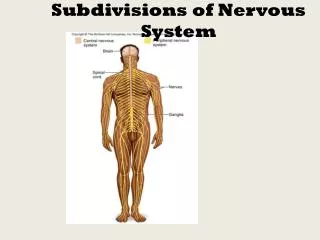
Somatic Sensory Pathways • carry sensory information from the skin and musculature of the body wall, head, neck, and limbs • posterior column pathway • anterolateral pathway • spinocerebellar pathway
The Posterior Column Pathway • Carries highly localizes fine touch, pressure, vibration, and proprioception • also called dorsal column/medial lemniscus First-Order Neuron (DRG, CN) 2 spinal tracts: • fasciculus gracilis • lower half of body • synapse in nucleus gracilis in medulla • fasciculus cuneatus • upper half of body • synapse in nucleus cuneatus in medulla
The Posterior Column Pathway Second-Order Neuron • soma in nucleus gracilis in medulla • soma in nucleus cuneatus in medulla • both ascend and cross over to the opposite side of the brain (decussation) and enter the medial lemniscus tract. • both synapse in the ventral nuclei of the thalamus
The Posterior Column Pathway Third-order Neuron • soma in ventral nuclei of the thalamus • arriving information is sorted • nature of stimulus • region of body involved • the axon travels to a specific region of the primary sensory cortex
Sensory Homunculus • electrical stimulation of human brains was used to create a functional map of the primary sensory cortex • the homunculus has a huge face because the face has the most sensory receptors and thus give the most sensory input
The Anterolateral Pathway • conscious, poorly localized sensations of touch, pressure, pain and temperature • first-order neurons • synapse on second-order neurons in the posterior gray horn of the spinal cord
The Anterolateral Pathway • second-order neurons (soma in spinal cord) • decussate • fibers ascend in • anterior spinothalamic tract • touch and pressure • lateral spinothalamic tract • pain and temperature • synapse on third-order neurons of thalamus
The Anterolateral Pathway • third-order neurons • soma in ventral nuclei of the thalamus • arriving information is sorted • nature of stimulus • region of body involved • the axon travels to a specific region of the primary sensory cortex
Phantom Limb Pain • An individual can experience painful sensations from an amputated limb, or if they were born without the limb. • The sensory neurons are still firing and the signal reaches the primary cortex and thus is perceived as a real stimulus on an absent limb.
Referred Pain • Perception of pain that is incorrectly localized. • Sensations arriving at segment of spinal cord can stimulate interneurons that are part of anterolateral pathway • heart attack • referred pain in left arm • appendicitis • referred pain in the naval and RLQ
The Spinocerebellar Pathway • proprioceptive input from muscles, tendons and joints • first-order neurons • synapse on second-order neurons in dorsal gray horn