Download
1 / 43
440 likes | 727 Views
Recognizing Different Sports Injuries. Chapter 9. Acute vs. Chronic Injuries. Acute Injuries New injuries Caused by trauma Physical injury or wound produced by internal or external force Chronic Injuries Result from overuse Happen over a period of time. Types of Synovial Joints.

E N D
Recognizing Different Sports Injuries Chapter 9
Acute vs. Chronic Injuries • Acute Injuries • New injuries • Caused by trauma • Physical injury or wound produced by internal or external force • Chronic Injuries • Result from overuse • Happen over a period of time
Types of Bones According to Shape • Long • Longer than wide • Help with movement • Short • Short and cube shaped • Carpals and tarsals • Flat • Flat and wide • protection • Irregular • Different sizes and functions
5 Functions of Bones • Provide shape and support • Allow movement • Give the muscles a place to attach • Protection • Protect internal organs • Store minerals • Calcium and phosphorus • Hematapoiesis (produce blood cells)
Parts of a Long Bone • Diaphysis – main shaft of a bone • Hollow, cylindrical, covered by compact bone • Epiphysis – ends of a bone • Growth area; “growth plate” • Articular cartilage – covers the end or epiphysis • Protection during movement, and cushions joint • Periosteum – dense, white membrane that covers diaphysis • Interlaced with muscle tendons, blood vessels, and osteoblasts (bone-forming cells)
Growth Plate Long shaft covering Lines joints
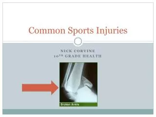
Fractures = breaks/cracks in bones Acute injuries Result of extreme stress and strains placed on the bone
Fractures (Fx) • Partial or complete tissue disruption that can be closed or open • Serious musculoskeletal injury • Signs/Symptoms: • Possible deformity, point tenderness, swelling, pain with both active and passive ROM • Forces • Tension, compression, bending, twisting, and shearing
Types of Acute Fractures • Greenstick Fx: • Incomplete break • Bone that have not ossified • Bones bend • Transverse Fx: • Straight line across bone • direct blow from the side • Spiral Fx: • “S” shaped • Foot is planted and body gets twisted
Types of Acute Fractures • Comminuted Fx: • Broken into 3 or more fragments • Hard blow or fall • Difficult to heal • Linear Fx: • Bone splints length wise • Landing from tall height or landing • Force applied to ends of bone • Oblique Fx: • Break is at an angle; similar to spiral • One end stabilized and the other twists
Video Clips Types of Fractures - 2 min How X-rays work - 2 min
Stress Fracture Chronic or overuse Fx Begins as dull ache and progressively gets worse; becomes very point tender Early on – pain during activity Late stages – pain after activity Common sites: tibia (shin) and metatarsals
Fracture Healing • Requires immobilization • Arms/legs: 6 weeks • Hands/feet: 3 weeks • Osteoblasts in the periostium form new bone cells as a callus on the fracture site • After immobilization normal stresses will help the bone re-model and heal
Dislocation When at least 1 bone of a joint is forced completely out of alignment or place Common: fingers, elbow, and shoulder Once dislocated = greater chance of dislocating again Shoulders most commonly dislocate in the anterior direction Sling and swathe – best way to immobilize “Seperated shoulder” is an AC joint sprain not a dislocation
Subluxation At least 1 bone comes partially out of its normal alignment or articulation and then goes back into place Common: shoulder and patella (kneecap) Need x-ray to rule out fx for dislocation and subluxation Return to play determined by soft tissue damage
Write the Question and Answer What is the difference in acute and chronic injuries? Draw a long bone and label the 4 parts. What are the 4 types of bones (classified by shape) and give an example of each? List the 5 functions of bones. What are the types of acute fractures? What is a stress fracture? What is the difference in a dislocation and subluxation?
Ligament Sprains • Ligaments – connect bone to bone • Gives support to a joint • Sprain – damage to a ligament • 3 grades of sprains • Grade 1 – some pain, no loss of function, mild pt. tenderness, mild swelling • Grade 2 – partial tearing of fibers and joint instability, pain, moderate loss of function • Grade 3 – complete tear, severe instability and swelling, extreme pain, and full loss of function
Ligament Damage Ligaments connect bone to bone Try to restore joint stability after a ligament injury Inelastic scar tissue forms, so ligament never regains the original tension it had Must increase strength around the joint
Contusion • Contusion = Bruise • From a direct blow • Tissue is compressed against object and the bone • Causes bleeding under the skin
MyositisOssificans • Calcium deposits form in the muscle • Tissue often compressed between object and bone • From repeated blows • When a muscle is bruised repeatedly • Quadriceps and biceps • Extremely painful and decreases ROM • Pad an area to prevent more bruising
Muscle Strain • Strain – stretch or tear of a muscle or tendon • Tendon – connects muscle to bone • 3 grades of strains • Grade 1 – some fibers stretched or torn. Movement it painful, but have full ROM • Grade 2 – more fibers torn; AROM extremely painful; feel depression or divot at tear; swelling and discoloration • Grade 3 – complete tear; possible total loss of AROM; extremely painful but goes away because nerves are severed or cut
Muscle Strains Grade 3 strain (rupture) Any muscle strain requires lengthy rehab Returning too early will re-injure the muscle
Muscle Guarding Involuntary muscle contractions that occur in response to a musculoskeletal injury Body’s way to splint the injury
Muscle Cramps • Painful involuntary contraction • Calf, hamstring, and abdomen • Often result of dehydration • Loss of water and electrolytes • Treatment • Stretch, replace water and electrolytes
Muscle Soreness • Muscular pain that is caused by overexertion during exercise • As a person gets older muscle soreness is more common • 2 Types • Acute-onset muscle soreness • Happens with muscle fatigue • During and just after exercise • Delayed-onset muscle soreness (DOMS) • Most intense 24-48 hours after exercise • Goes away 3-4 days later • Stretch and ice
Inflammation • Usually thought of as bad, but is an essential part of the healing process • Once an structure is damaged, inflammation must occur to start the healing process • Signs and Symptoms • Pain, swelling, warmth, redness, loss of function • -itis means inflammation
Tendonitis • Inflammation of a tendon • Most common overuse injury • Pain, swelling, maybe warmth and crepitus • Crepitus – crackling feeling or sound • Treatment • REST or at least change activities to decrease stress • Tendon sticks to surrounding structures when moving
Tenosynovitis Inflammation of a tendon and the synovial sheath (or covering) it slides through Sheath is present to reduce friction and help movement Tendinitis makes the tendon “sticky” so it becomes difficult to move through the sheath REST, change activities, medications, ice
Bursitis Inflammation of a bursae Bursae – fluid filled sac at joints that help with movement and provide protection When inflammed the bursae produces more fluid which makes the space crowded and produces pain REST, change activities, medications, ice
Neuritis • Inflammation of a nerve • S&S of a nerve injury • Numbness, tingling, loss of sensation, burning • Common Nerve injuries • “Stinger” – brachial plexus is stretched in shoulder • “funny bone” – ulnar nerve is hit in elbow
The Healing Process • Occurs after all injuries • Can’t speed up the healing process, but things can be done to slow it down • Consists of 3 phases: • Inflammatory response phase • Fibroblastic repair phase • Maturation-remodeling phase • The phases overlap - continuum
1. Inflammatory Response Phase Begins as soon as injury occurs Inflammation must occur or other phases won’t Most critical part The damaged cells are “cleaned up” Damaged cells release chemicals that will help healing later Lasts approximately 2-4 days following the injury
2. Fibroblastic Repair Phase Regenerative activity leads scar formation Scar formation begins a few hours after the injury and lasts for 4-6 weeks Signs and symptoms of inflammation go away As scar formation progresses tenderness and pain go away
3. Maturation-Remodeling Phase Long term process The scar remodels and becomes stronger as it changes shape Collagen fibers of scar are parallel to forces Tissue has normal appearance, but scar is not as strong as uninjured tissue Strong scar is present in about 3 weeks Begins 2-3 weeks after injury and lasts for several years