Download
1 / 56
580 likes | 768 Views
Chapter 27. Chest and Abdominal Trauma. Anatomy and Physiology of the Chest. Anatomy and Physiology of the Chest. Thoracic Cavity. Subdivided into two smaller spaces Mediastinum – in center Contains heart, great vessels, esophagus, trachea, nerves

E N D
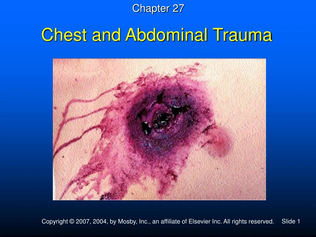
Chapter 27 Chest and Abdominal Trauma
Thoracic Cavity • Subdivided into two smaller spaces • Mediastinum – in center • Contains heart, great vessels, esophagus, trachea, nerves • Pleural spaces – on either side of mediastinum
Chest Trauma – Mechanisms of injury • Blunt • Sudden deceleration of chest wall against a fixed object • Penetration
Rib Fractures • Most often the result of blunt trauma • Isolated rib fracture usually not a serious emergency • Can puncture lung or blood vessel • Pneumothorax, hemothorax, flail chest • Lower rib fractures may injure abdominal organs • Liver, spleen, kidneys
Case History You respond to an MVC to find a 65-year-old female victim of a front end collision. She is complaining of severe chest pain and dyspnea. She is pale, cyanotic, and diaphoretic. You notice that the steering wheel is deformed.
Flail Chest • Two or more ribs fractured in two or more places • Paradoxical chest movement • Look for signs of underlying injury (e.g., pneumothorax)
Flail Chest – Management • Splint chest wall • Blanket, towel, sheet • Rigid splint • Positive-pressure ventilation • When hypoventilation is present and patient can tolerate • Restores adequate ventilation • Otherwise nonrebreather device
Traumatic Asphyxia • Severe compression of thorax • High-velocity or steering wheel injuries, heavy weight dropped on chest • Heart compressed; blood driven to thorax and neck • Ecchymosis and edema • Life-threatening injury • Look for associated injuries to lungs and chest wall • Management: high-concentration oxygen, possible PPV
Case History You respond to a call for “difficulty breathing” to find a 19-year-old male complaining of dyspnea and chest pain. He states that it started suddenly while he was running. His breathing difficulty has gotten worse over the last hour.
Pneumothorax • Occurs when air enters visceral and parietal pleura • Collapses lung • Less alveolar surface for diffusion of oxygen • Results in hypoxia • Two mechanisms • Trauma • Spontaneous rupture
Traumatic • Penetrating • Missile • Sharp object • Broken rib • Blunt • Person takes deep breath just before auto collision • “Paper bag effect”
Spontaneous • Ruptured bleb in lung tissue • Young, muscular males • COPD patients
Open Pneumothorax – Assessment and Recognition • Sucking wound • Dyspnea • Pleuritic chest pain
Open Pneumothorax – Assessment and Recognition • Absent or diminished breath sounds on affected side • Signs of respiratory distress • Subcutaneous emphysema • Historical profile
Open Pneumothorax – Management • Check ABCs. • Administer oxygen; positive-pressure ventilation, if needed (carefully). • Seal wound with airtight dressing on three sides. • Place patient in position of comfort. • Transport to definitive care.
Closed Pneumothorax • Also called simple pneumothorax • Management • High-concentration oxygen; possible PPV • Transport without delay. • Watch for signs of a developing tension pneumothorax.
Case History You respond to an MVC and find a 32-year-old female involved in victim of a front end collision complaining of severe chest pain and dyspnea. She is pale, cyanotic, and diaphoretic. The police on scene says she was fine when they arrived but she suddenly started to become “very sick.”
Tension Pneumothorax • Air trapped within pleural space • Acts as a one-way valve • Increased intrathoracic pressure • Can collapse superior and inferior vena cavae • Reduces blood return to heart • Causes profound shock
Tension Pneumothorax – Assessment and Recognition • Increasing respiratory distress and cyanosis • Breath sounds absent on affected side • Distended neck veins • Tracheal shift • Signs of shock
Tension Pneumothorax – Management • If airtight dressing was applied, remove dressing • Reapply dressing after air escapes. • Watch for further tension. • Transport immediately. • Consider ALS intercept (for needle decompression).
Hemothorax • Blood within the pleural space • Thorax has the capacity for massive blood loss. • Physiologic effects • Primary effect – hypovolemic shock • May exist with or without an associated pneumothorax • May occur due to penetrating injuries or to rib fractures
Hemothorax – Assessment and Recognition • Signs of hypovolemic shock • Delayed or absent capillary refill (children) • Pale, cool, sweaty skin • Tachycardia • Rapid and shallow breathing
Hemothorax – Assessment and Recognition • Breath sounds absent on the affected side • Hemoptysis (coughing blood) • Hypotension (late sign) • Altered mental state (late sign) • Cardiovascular collapse (cardiac arrest)
Hemothorax – Management • Establish a patent airway. • Suction available to manage hemoptysis • High-concentration oxygen; possible PPV • Transport immediately.
Pulmonary Contusion • From severe blows to chest wall • Can result in swelling and fluid buildup • Decreases diffusion of oxygen into capillaries • Management • High-concentration oxygen
Cardiac Tamponade • Fluid accumulation in the pericardial sac caused by bleeding or fluid loss • May result from blunt or penetrating trauma
Cardiac Tamponade — Physiologic Effects • Bleeding places pressure on atria, ventricles, and vena cava. • Venous return is obstructed. • Interferes with the normal dynamics of contraction. • Cardiac output is decreased.
Cardiac Tamponade — Assessment and Recognition • Penetrating wound or precordial contusion may be present. • Signs of shock • Decreased pulse pressure • Muffled heart sounds • Distended neck veins
Cardiac Tamponade — Management • Early recognition and rapid hospital intervention – most essential
Case History You respond to a “man down” to find a 20-year-old construction worker who fell 30 feet from a rooftop. He is complaining of pain in his chest and back. He is pale and diaphoretic. His pulse is 130 and thready. He is responsive to painful but not verbal stimuli.
Aortic Tear • Complete tear results in exsanguination and death. • Partial tear causes leak and hemorrhage. • Hypovolemic shock is main problem. • Mortality is very high from massive hemorrhage. • 80% die within first hour
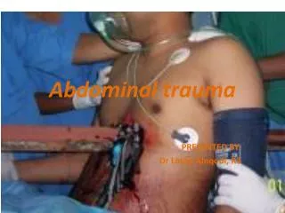
Abdominal Injuries • Large vessels and highly vascular organs within abdomen • Rapid blood loss and death • Maintain high level of suspicion • May be from blunt or penetrating trauma • Primary goal • Recognize life-threatening injuries. • Administer essential life support. • Transport without delay (requires surgical intervention).
Mechanism of Injury • Blunt trauma • Compression injuries • Deceleration injuries • Seat belt injuries • Penetrating trauma
Assessment • Scene size-up • Obtain MOI • Initial assessment • Look for signs of hypovolemia
Assessment • Focused history and physical examination • Look for bruises, tire marks, seat belt marks. • Is abdomen distended? • DCAP-BTLS • Palpate abdomen for tenderness and guarding. • Save painful area for last. • Palpate iliac crest. • If pelvic bones move, stop examination.
Assessment • Focused history and physical examination (continued) • Associated head or spinal injuries may present with loss of pain perception. • SAMPLE history • Elderly? • History of medications that slow heart rate? • Signs of alcohol or drugs
Management • Management occurs in hospital. • Treat for shock. • Transport without delay.
Special Considerations • Evisceration • Do not attempt to put organs back in abdomen. • Cover with moist, sterile dressing or airtight dressing. • Transport in supine position with hips and legs flexed with pillow under knees.
Special Considerations • Urinary tract injuries • Look for bruises over flank. • Injuries to pelvis can cause bladder or urethral tears. • Injuries to male genitalia • May result in lacerations, bruising, avulsion, or amputation • Injuries to female genitalia • May occur from direct trauma or straddle injuries
Acute Abdomen • Recent onset of abdominal pain • Requires early diagnosis and surgical intervention