Download
1 / 107
1.07k likes | 1.11k Views
DNA: The Genetic Material. Chapter 16 Molecular Basis of Inheritance. The Genetic Material. Frederick Griffith, 1928 studied Streptococcus pneumoniae , a pathogenic bacterium causing pneumonia there are 2 strains of Streptococcus : - S strain is virulent - R strain is nonvirulent

E N D
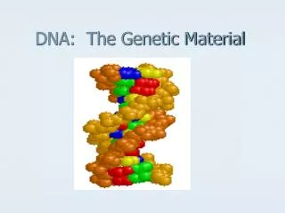
DNA: The Genetic Material Chapter 16 Molecular Basis of Inheritance
The Genetic Material Frederick Griffith, 1928 studied Streptococcus pneumoniae, a pathogenic bacterium causing pneumonia there are 2 strains of Streptococcus: - S strain is virulent - R strain is nonvirulent Griffith infected mice with these strains hoping to understand the difference between the strains
The Genetic Material Griffith’s results: - live S strain cells killed the mice - live R strain cells did not kill the mice - heat-killed S strain cells did not kill the mice - heat-killed S strain + live R strain cells killed the mice
The Genetic Material Griffith’s conclusion: - information specifying virulence passed from the dead S strain cells into the live R strain cells - Griffith called the transfer of this information transformation
The Genetic Material Avery, MacLeod, & McCarty, 1944 Repeated Griffith’s experiment using purified cell extracts and discovered: - removal of all protein from the transforming material did not destroy its ability to transform R strain cells - DNA-digesting enzymes destroyed all transforming ability - the transforming material is DNA
The Genetic Material Hershey & Chase, 1952 - investigated bacteriophages: viruses that infect bacteria - the bacteriophage was composed of only DNA and protein - they wanted to determine which of these molecules is the genetic material that is injected into the bacteria
The Genetic Material - BacteriophageDNAwas labeled with radioactive phosphorus (32P) - Bacteriophage protein was labeled with radioactive sulfur (35S) - radioactive molecules were tracked - only the bacteriophage DNA (as indicated by the 32P) entered the bacteria and was used to produce more bacteriophage - conclusion: DNA is the genetic material
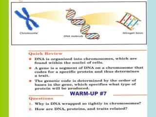
DNA Structure Determining the 3-dimmensional structure of DNA involved the work of a few scientists: • Erwin Chargaff determined that • amount of adenine = amount of thymine • amount of cytosine = amount of guanine This is known as Chargaff’s Rules
DNA Structure DNA is a nucleic acid. The building blocks of DNA are nucleotides, each composed of: • a 5-carbon sugar called deoxyribose • a phosphate group (PO4) • a nitrogenous base • adenine, thymine, cytosine, guanine
Albrecht Kossel found Bases in 1880s – 1890s 1920s Phoebus Levene discovered Nucleotides: Sugar and Phosphates that bond together. Phosphodiester Bonds
DNA Structure Nucleotides are connected to each other to form a long chain phosphodiester bond: bond between adjacent nucleotides • formed between the phosphate group of one nucleotide and the 3’ –OH of the next nucleotide The chain of nucleotides has a 5’ to 3’ orientation.
DNA Structure The nucleotide structure consists of • the nitrogenous base attached to the 1’ carbon of deoxyribose • the phosphate group attached to the 5’ carbon of deoxyribose • a free hydroxyl group (-OH) at the 3’ carbon of deoxyribose
DNA Structure Rosalind Franklin and Maurice Wilkins • Franklin performed X-ray diffraction studies to identify the 3-D structure • discovered that DNA is helical • discovered that the molecule has a diameter of 2nm and makes a complete turn of the helix every 3.4 nm
DNA Structure James Watson and Francis Crick, 1953 • deduced the structure of DNA using evidence from Chargaff, Franklin, and others • proposed a double helixstructure
DNA is a Double Helix • Sugar and phosphate form the backbone • covalently bonded to each other ("phosphodiester bonds") • Bases lie between the backbone • Nucleotides • Held together by H-bonds between the complimentary bases • A-T – 2 H bonds • G-C – 3 H bonds
DNA Structure The two strands of nucleotides are antiparallel to each other • one is oriented 5’ to 3’, the other 3’ to 5’ The two strands wrap around each other to create the helical shape of the molecule.
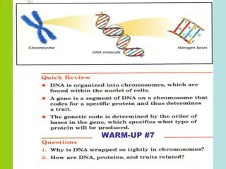
Orientation of DNA • Phosphate end is the 5' end • The opposite end (sugar) is the 3' end • Q: Since DNA is complementary, what end matches with the 5’ end?
DNA Replication Matthew Meselson & Franklin Stahl, 1958 investigated the process of DNA replication considered 3 possible mechanisms: • conservative model • semiconservative model • dispersive model
DNA Replication Meselson and Stahl concluded that the mechanism of DNA replication is the semiconservative model. Each strand of DNA acts as a template for the synthesis of a new strand.
Semiconservative Replication = each strand has 1 parent and 1 new strand
Replication • DNA Helicase unzips DNA • RNA Primers bind to DNA strands • DNA Polymerase adds nucleotides to DNA • Leading – continuous adding of bases • Lagging – Okazaki fragments • DNA Ligase fills in gaps
3’ Parental DNA Molecule 5’ Replication Fork 3’ 5’ DNA Replication • Two strands open forming Replication Forks (Y-shaped region) • New strands grow at the forks
Unwinding • DNA Helicase unwinds/unzips DNA strand • Hydrogen bonds are broken • RNA Primase adds a starter segment to each strand
Base Pairing Steps • DNA Polymerase adds nucleotides to DNA strands • DNA polymerase can only add nucleotides to the 3’ end of the DNA • This causes the NEW strand to be built in a 5’ to 3’ direction • Adds a 5’ DNA polymerase III (pol III) is the main polymerase responsible for the majority of DNA synthesis -Adds nucleotides to the 3’ end of the daughter strand of DNA
Leading Strand Replicated as the DNA unwinds New nucleotides are added continuously
Joining DNA polymerase removes RNA primer and fills with DNA nucleotide DNA Ligase – links two sections of DNA together
Lagging Strand Replicated away from replication fork Create segments - Okazaki Fragment Fragments are later connected
3’ 5’ Which Template will be the leading strand? Lagging strand? 5’ 3’ What would be the complementary DNA strand for the following DNA sequence? DNA 5’-CGTATG-3’
SWEET VID!! https://www.youtube.com/watch?v=TEQMeP9GG6M
Prokaryote vs. Eukaryote Prokaryotes • 1 origin Eukaryote • Many origins
Eukaryotic DNA Replication The larger size and complex packaging of eukaryotic chromosomes means they must be replicated from multiple origins of replication. The enzymes of eukaryotic DNA replication are more complex than those of prokaryotic cells.
Eukaryotic DNA Replication Synthesizing the ends of the chromosomes is difficult because of the lack of a primer. With each round of DNA replication, the linear eukaryotic chromosome becomes shorter.
Eukaryotic DNA Replication telomeres – repeated DNA sequence on the ends of eukaryotic chromosomes • produced by telomerase telomerase contains an RNA region that is used as a template so a DNA primer can be produced
Telomeres 1. Each round of replication shortens the 5' end of the lagging strand (by about 100-200 bp) 2. If this continued indefinitely, chromosomes would get shorter and shorter after each replication. • Information would start to be lost 3. TELOMERASE - enzyme responsible for adding telomeres at end of eukaryotic chromosomes a. short, repeating DNA sequence
DNA Repair - DNA-damaging agents - repair mechanisms - specific vs. nonspecific mechanisms
DNA Repair Mistakes during DNA replication can lead to changes in the DNA sequence and DNA damage. DNA can also be damaged by chemical or physical agents called mutagens. Repair mechanisms may be used to correct these problems.