Download
1 / 204
2.08k likes | 2.35k Views
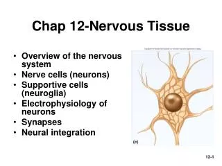
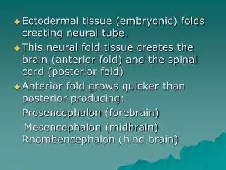
12 Neural Tissue. An Introduction to the Nervous System. Learning Outcomes 12-1 Describe the anatomical and functional divisions of the nervous system.

E N D
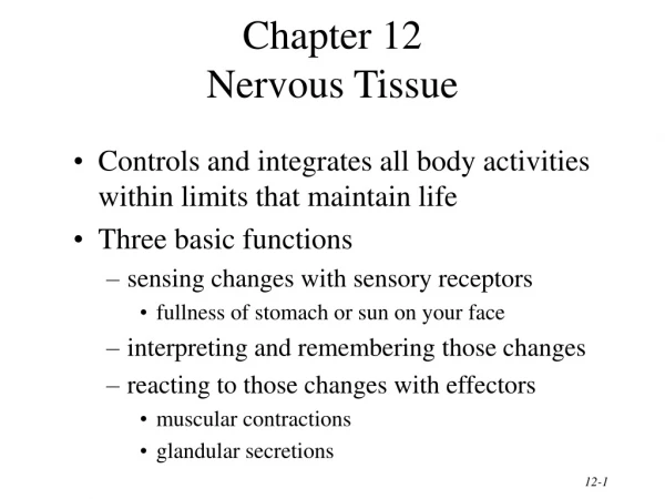
12 Neural Tissue
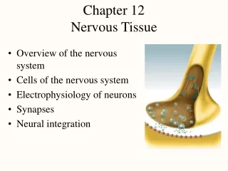
An Introduction to the Nervous System • Learning Outcomes • 12-1 Describe the anatomical and functional divisions of the nervous system. • 12-2 Sketch and label the structure of a typical neuron, describe the functions of each component, and classify neurons on the basis of their structure and function. • 12-3 Describe the locations and functions of the various types of neuroglia.
An Introduction to the Nervous System • Learning Outcomes • 12-4 Explain how the resting potential is created and maintained. • 12-5 Describe the events involved in the generation and propagation of an action potential. • 12-6 Discuss the factors that affect the speed with which action potentials are propagated.
An Introduction to the Nervous System • Learning Outcomes • 12-7 Describe the structure of a synapse, and explain the mechanism involved in synaptic activity. • 12-8 Describe the major types of neurotransmitters and neuromodulators, and discuss their effects on postsynaptic membranes. • 12-9 Discuss the interactions that enable information processing to occur in neural tissue.
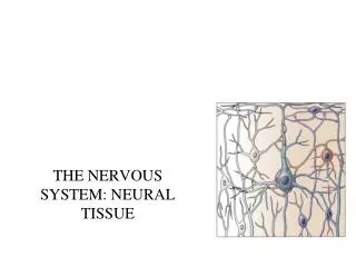
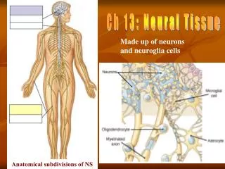
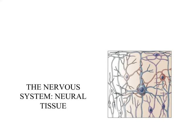
An Introduction to the Nervous System • The Nervous System • Includes all neural tissue in the body • Neural tissue contains two kinds of cells • Neurons • Cells that send and receive signals • Neuroglia (glial cells) • Cells that support and protect neurons
An Introduction to the Nervous System • Organs of the Nervous System • Brain and spinal cord • Sensory receptors of sense organs (eyes, ears, etc.) • Nerves connect nervous system with other systems
12-1 DivisionsoftheNervousSystem • Anatomical Divisions of the Nervous System • Central nervous system (CNS) • Peripheral nervous system (PNS)
12-1 DivisionsoftheNervousSystem • The Central Nervous System (CNS) • Consists of the spinal cord and brain • Contains neural tissue, connective tissues, and blood vessels • Functions of the CNS are to process and coordinate: • Sensory data from inside and outside body • Motor commands control activities of peripheral organs (e.g., skeletal muscles) • Higher functions of brain intelligence, memory, learning, emotion
12-1 DivisionsoftheNervousSystem • The Peripheral Nervous System(PNS) • Includes all neural tissue outside the CNS • Functions of the PNS • Deliver sensory information to the CNS • Carry motor commands to peripheral tissues and systems
12-1 DivisionsoftheNervousSystem • The Peripheral Nervous System (PNS) • Nerves (also called peripheral nerves) • Bundles of axons with connective tissues and blood vessels • Carry sensory information and motor commands in PNS • Cranial nerves — connect to brain • Spinal nerves — attach to spinal cord
12-1 DivisionsoftheNervousSystem • Functional Divisions of the PNS • Afferent division • Carries sensory information • From PNS sensory receptorsto CNS • Efferent division • Carries motor commands • From CNS to PNS muscles and glands
12-1 DivisionsoftheNervousSystem • Functional Divisions of the PNS • Receptors and effectors of afferent division • Receptors • Detect changes or respond to stimuli • Neurons and specialized cells • Complex sensory organs (e.g., eyes, ears) • Effectors • Respond to efferent signals • Cells and organs
12-1 DivisionsoftheNervousSystem • Functional Divisions of the PNS • The efferent division • Somatic nervous system(SNS) • Controls voluntary and involuntary (reflexes) muscle skeletal contractions
12-1 DivisionsoftheNervousSystem • Functional Divisions of the PNS • The efferent division • Autonomic nervous system (ANS) • Controls subconscious actions, contractions of smooth muscle and cardiac muscle, and glandular secretions • Sympathetic division has a stimulating effect • Parasympathetic division has a relaxing effect
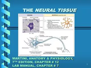
12-2 Neurons • Neurons • The basic functional units of the nervous system • The structure of neurons • The multipolar neuron • Common in the CNS • Cell body (soma) • Short, branched dendrites • Long, single axon
12-2 Neurons • The Cell Body • Large nucleus and nucleolus • Perikaryon (cytoplasm) • Mitochondria (produce energy) • RER and ribosomes (produce neurotransmitters)
12-2 Neurons • TheCell Body • Cytoskeleton • Neurofilaments and neurotubules in place of microfilaments and microtubules • Neurofibrils: bundles of neurofilaments that provide support for dendrites and axon • Nissl bodies • Dense areas of RER and ribosomes • Make neural tissue appear gray (gray matter)
12-2 Neurons • Dendrites • Highly branched • Dendritic spines • Many fine processes • Receive information from other neurons • 80–90% of neuron surface area
12-2 Neurons • The axon • Is long • Carries electrical signal (action potential) to target • Axon structure is critical to function
12-2 Neurons • Structures of the Axon • Axoplasm • Cytoplasm of axon • Contains neurofibrils, neurotubules, enzymes, organelles • Axolemma • Specialized cell membrane • Covers the axoplasm
12-2 Neurons • Structures of the Axon • Axon hillock • Thick section of cell body • Attaches to initial segment • Initial segment • Attaches to axon hillock
12-2 Neurons • Structures of the Axon • Collaterals • Branches of a single axon • Telodendria • Fine extensions of distal axon • Synaptic terminals • Tips of telodendria
Figure 12-1a The Anatomy of a Multipolar Neuron Dendrites Perikaryon Cell body Nucleus Telodendria Axon This color-coded figure shows the four general regions of a neuron.
Figure 12-1b The Anatomy of a Multipolar Neuron Dendritic branches Nissl bodies (RER and free ribosomes) Mitochondrion Axon hillock Initial segment of axon Axolemma Telodendria Axon Golgi apparatus Neurofilament Synaptic terminals Nucleus Nucleolus See Figure 12–2 Dendrite An understanding of neuron function requires knowing its structural components. PRESYNAPTIC CELL POSTSYNAPTIC CELL
12-2 Neurons • The Structure of Neurons • The synapse • Area where a neuron communicates with another cell
12-2 Neurons • The Structure of Neurons • The synapse • Presynaptic cell • Neuron that sends message • Postsynaptic cell • Cell that receives message • The synaptic cleft • The small gap that separates the presynaptic membrane and the postsynaptic membrane
12-2 Neurons • The Synapse • The synaptic terminal • Is expanded area of axon of presynaptic neuron • Contains synaptic vesicles of neurotransmitters
12-2 Neurons • Neurotransmitters • Are chemical messengers • Are released at presynaptic membrane • Affect receptors of postsynaptic membrane • Are broken down by enzymes • Are reassembled at synaptic terminal
12-2 Neurons • Recycling Neurotransmitters • Axoplasmic transport • Neurotubules within the axon • Transport raw materials • Between cell body and synaptic terminal • Powered by mitochondria, kinesin, and dynein
12-2 Neurons • Types of Synapses • Neuromuscular junction • Synapse between neuron and muscle • Neuroglandular junction • Synapse between neuron and gland
Figure 12-2 The Structure of a Typical Synapse Telodendrion Synaptic terminal Endoplasmic reticulum Mitochondrion Synaptic vesicles Presynaptic membrane Synaptic cleft Postsynaptic membrane
12-2 Neurons • Structural Classification of Neurons • Anaxonic neurons • Found in brain and sense organs • Bipolar neurons • Found in special sensory organs (sight, smell, hearing) • Unipolar neurons • Found in sensory neurons of PNS • Multipolar neurons • Common in the CNS • Include all skeletal muscle motor neurons
12-2 Neurons Anaxonic Neurons Small All cell processes look alike Bipolar Neurons Are small One dendrite, one axon
12-2 Neurons Unipolar Neurons Also calledpseudounipolar neurons Have very long axons Fused dendrites and axon Cell body to one side Multipolar Neurons Have very long axons Multiple dendrites, one axon
Figure 12-3 A Structural Classification of Neurons Anaxonic neuron Bipolar neuron Unipolar neuron Multipolar neuron Dendrites Dendrites Initial segment Cell body Dendritic branches Axon Dendrite Cell body Cell body Axon Axon Cell body Axon Synaptic terminals Synaptic terminals Synaptic terminals
Figure 12-3a A Structural Classification of Neurons Anaxonic neuron Cell body Anaxonic neurons have more than two processes, but axons cannot be distinguished from dendrites.
Figure 12-3b A Structural Classification of Neurons Bipolar neuron Dendritic branches Dendrite Cell body Axon Synaptic terminals Bipolar neurons have two processes separated by the cell body.
Figure 12-3c A Structural Classification of Neurons Unipolar neuron Dendrites Initial segment Axon Cell body Axon Synaptic terminals Unipolar neurons have a single elongate process, with the cell body situated off to the side.
Figure 12-3d A Structural Classification of Neurons Multipolar neuron Dendrites Cell body Axon Synaptic terminals Multipolar neurons have more than two processes; there is a single axon and multiple dendrites.
12-2 Neurons • Three Functional Classifications of Neurons • Sensory neurons • Afferent neurons of PNS • Motor neurons • Efferent neurons of PNS • Interneurons • Association neurons
12-2 Neurons • Functions of Sensory Neurons • Monitor internal environment (visceral sensory neurons) • Monitor effects of external environment (somatic sensory neurons) • Structures of Sensory Neurons • Unipolar • Cell bodies grouped in sensory ganglia • Processes (afferent fibers) extend from sensory receptors to CNS
12-2 Neurons • Three Types of Sensory Receptors • Interoceptors • Monitor internal systems (digestive, respiratory, cardiovascular, urinary, reproductive) • Internal senses (taste, deep pressure, pain) • Exteroceptors • External senses (touch, temperature, pressure) • Distance senses (sight, smell, hearing) • Proprioceptors • Monitor position and movement (skeletal muscles and joints)
12-2 Neurons • Motor Neurons • Carry instructions from CNS to peripheral effectors • Via efferent fibers (axons)
12-2 Neurons • Motor Neurons • Two major efferent systems • Somatic nervous system (SNS) • Includes all somatic motor neurons that innervate skeletal muscles • Autonomic (visceral) nervous system (ANS) • Visceral motor neurons innervate all other peripheral effectors • Smooth muscle, cardiac muscle, glands, adipose tissue
12-2 Neurons • Motor Neurons • Two groups of efferent axons • Signals from CNS motor neurons to visceral effectors pass synapses at autonomic ganglia dividing axons into: • Preganglionic fibers • Postganglionic fibers
12-2 Neurons • Interneurons • Most are located in brain, spinal cord, and autonomic ganglia • Between sensory and motor neurons • Are responsible for: • Distribution of sensory information • Coordination of motor activity • Are involved in higher functions • Memory, planning, learning
12-3 Neuroglia • Neuroglia • Half the volume of the nervous system • Many types of neuroglia in CNS and PNS
12-3 Neuroglia • Four Types of Neuroglia in the CNS • Ependymal cells • Cells withhighly branched processes; contact neuroglia directly • Astrocytes • Large cell bodies with many processes • Oligodendrocytes • Smaller cell bodies with fewer processes • Microglia • Smallest and least numerous neuroglia with many fine-branched processes
12-3 Neuroglia • Ependymal Cells • Form epithelium called ependyma • Line central canal of spinal cord and ventricles of brain • Secrete cerebrospinal fluid (CSF) • Have cilia or microvilli that circulate CSF • Monitor CSF • Contain stem cells for repair
12-3 Neuroglia • Astrocytes • Maintain blood–brain barrier (isolates CNS) • Create three-dimensional framework for CNS • Repair damaged neural tissue • Guide neuron development • Control interstitial environment