Download
1 / 33
340 likes | 536 Views
The Nervous System: Neural Tissue. Coordination of activities: Nervous System. Nervous System: Swift, brief, temporary Functions: (motor & sensory) Provide sensation Integrate info Coordinate voluntary & involuntary activities Regulate & control systems. Nervous System Overview.

E N D
Coordination of activities:Nervous System • Nervous System: • Swift, brief, temporary • Functions: (motor & sensory) • Provide sensation • Integrate info • Coordinate voluntary & involuntary activities • Regulate & control systems
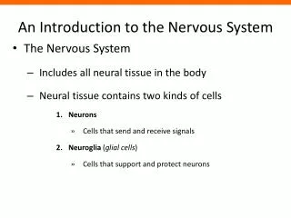
Nervous System Overview • 2 Anatomical Divisions • Central Nervous System (CNS) • Brain and spinal cord • Peripheral Nervous System (PNS) • Somatic Nervous System (SNS) • Autonomic Nervous System (ANS) • Sympathetic N. S.- “flight or fight” • Parasympathetic N. S.- “rest & repose” • http://itc.gsw.edu/faculty/gfisk/anim/index.html • 2 Functional Divisions (based on function) • Afferent: Sensory (muscles/ glands CNS) • Efferent: Motor (CNS muscles/ glands
Cellular Organization in Neural Tissue • Neurons: (=Nerve cells) • Soma (cell body) • Dendrites • Axon & Nodes of Ranvier/ Internodes • Synaptic terminals (knobs) • Neuroglia= Glial Cells • Protects neurons • Provides support • Phagocytes
Neuron Functional Classifications • Sensory Neurons= Afferent fibers • TO CNS from ext & int environment • Cell bodies in peripheral ganglia • neurons • from sensory receptors spinal cord • Types: • Somatic Sensory Neurons: ext environment • Exteroceptors: sight, smell, hear, touch • Proprioceptors: position joints & muscles • Visceral Sensory Neurons= Interoceptors: internal envmt like visceral systems & taste, deep pressure, pain
Neuron Functional Classification • Motor Neurons: Efferent fibers • From CNS to effectors (muscles/ glands) • Stimulates or modifies activity of peripheral tissues, organs, organ systems • Types: • Somatic Motor Neurons: • Voluntary skeletal muscle • Spinal cord neuromuscular junction • Visceral Motor Neurons (of ANS) • Involuntary peripheral effectors • Spinal Cord (preganglionic fibers) peripheral ganglion (PNS cell bodies) postganglionic fibers effectors
Neuron Functional Classification • Interneurons= Association Neurons • b/w sensory & motor neurons • Located entirely in brain & spinal cord • Function: • Analysis of sensory inputs • Coordination of motor outputs • Classified as excitatory or inhibitory
Neuron Communication • Synapse • Space between synaptic knob & next cell • Neurons communicate with: • Other neurons • Other Cells: • Neuromuscular Junction • Neuroglandular Junction • Neurotransmitters • Chemical messenger at synapse
Resting Membrane Potential • Intracellular Fluids: High in [K+] • Extracellular Fluids: High in [Na+] • Na+/K+ exchange pump • Pump 2K+ in and 3 Na+ out • Resting potential: -70mV • Ion Channels: • Passive channels: always open & “leaky” • Active or Gated Channels: • Chemically regulated: neurotransmitter • Voltage regulated: voltage changes • Mechanically regulated: physical stim (Merkel’s discs)
Neuron Electrical Signal: Action Potential • Action Potential: • Electrical signal • Stimulated by neurotransmitter, voltage change, or physical stimulus • Rules to occur: • Depolarization to threshold (-60 to -55mV) • Occurs in axons & muscle fibers (down T-tubules) • All action potentials generated are identical (gun) • All-or-None Response (toll booth)
Action Potential Generation: Steps • Activate voltage-gated Na+ channelsdepolarization • Increase membrane potential: Na+ in • Inactivate Voltage-gated Na+ channels: • +30mV close & can’t be opened • Activate Voltage-Gated K+ channels repolarization • Decrease membrane potential (-70mV): K+ out • Return to resting membrane potential: -70mV • Voltage-gated Na+ channels closed, can open. • Voltage-gated K+ channels closing with delay of 1msec hyperpolarization (below -70mV) • After 1msec normal resting potential (-70mV) • Action Potential travels to next portion of membrane
Refractory Periods of Action Potentials • Absolute refractory period: Repolarization • Membrane CAN’T respond to stim • From opening voltage-gated Na+ channels end of action potential • Action potential: one direction away from soma. • Relative refractory period: Hyperpolarization • Only LARGER stimulus can initiate axn potential • From when voltage-gated Na+ channels are restored to normal (closed but able to open) state, but voltage-gated K+ channels still open.
Action Potential Conduction Velocity • Myelin sheath • Continuous conduction: segment by segment • Unmyelinated axons • Saltatory Conduction: leap from node to node • Myelinated axons: ions can’t pass lipid nodes • http://www.blackwellpublishing.com/matthews/actionp.html • Axon Diameter • Type A: Largest, myelinated fastest (300mph) • sk. mm. motor neurons, balance & L.T. • Type B: Smaller, myelinated medium (40mph) • Type C: Smallest, unmyelinated slowest (2mph) • Both B & C carry pain, temp, to smooth mm & glands
Other Factors on Rate of A.P. • Hormones can promote facilitation or inhibition • Presynaptic inhibition: inhibits Ca2+ channels decr postsynaptic stim (i.e. GABA) • Presynaptic Facilitation: keep Ca2+ channels open • [Na+, K+, Ca2+] (dehydration or kidney dz), pH (basic convulsions, acidic inhibition), temp changes
ATP needed for: Synthesis, release, & recycling NT Movement of materials Recovery from a.p.: [ion]- Na+/ K+ pump ATP Production: Aerobic glycolysis (no glucose stores) Need uninterrupted blood supply for O2 & glucose Stroke: less than 1-2 hrs few weeks to recover longer permanent ATP Needs & Production
Drug Effects: Interferes w/ NT synthesis Alter rate of NT release Prevent NT inactivation Prevent NT binding to receptors Caffeine lowers threshold at axon hillock more sensitive to stim (facilitation) “jumpy” Nicotine bind to receptor sites on postsynaptic membrane & stimulate it no enzymes to remove it Examples Botulinus toxin: blocks release Ach at presynaptic membrane paralysis Black Widow spiders venom : cause massive release of Ach cramps/ spasms Anticholinesterase drugs: block breakdown Ach by AchE extreme contract (ex: nerve gases, pst controls) Atropine: prevent Ach binding to postsynaptic neuron paralysis (curare plant extract) Ach Drugs & Synaptic Function