Download

1 / 29
310 likes | 669 Views
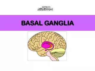
The Basal Ganglia. What are the basal ganglia?. Other Terms: Archistriatum Paleostriatum Neostriatum. Dorsal Striatum Caudate nucleus Putamen Ventral Striatum Nucleus Accumbens Olfactory Tubercle Globus Pallidus Internal segment External segment Ventral pallidum Subthalamic nucleus

E N D
What are the basal ganglia? Other Terms: Archistriatum Paleostriatum Neostriatum • Dorsal Striatum • Caudate nucleus • Putamen • Ventral Striatum • Nucleus Accumbens • Olfactory Tubercle • Globus Pallidus • Internal segment • External segment • Ventral pallidum • Subthalamic nucleus • Substantia nigra • Pars compacta • Pars reticulata • Pedunculopontine nucleus**
Caudate Nucleus • C shaped structure (“tail”) • Lateral wall of lateral ventricle • Head, body and tail
Caudate nucleus • Putamen • Nucleus accumbens • Internal capsule • External capsule • Extreme capsule • claustrum • Septum pellucidum • Insular cortex • Corpus callosum
Caudate nucleus • Putamen • Globus pallidus external • Globus pallidus internal • Ventral pallidum • Anterior commissure • Substantia innominata • Internal capsule • Lentiform nucleus**
Head, body, tail of caudate • anterior and temporal horn of lateral ventricle • Globus pallidus internal and external • Internal capsule, anterior and posterior limbs
Caudate nucleus (body and tail) • Putamen • Globus pallidus • Subthalamic nucleus • Substantia nigra • Pars compacta • Pars reticulata
Globus pallidus external • Globus pallidus internal • Subthalamic nucleus • Substantia nigra
Subthalamic nucleus • Substantia nigra • Ventral tegmental area
Globus pallidus and entopeduncular nucleus vs. Globus pallidus (external) and Globus pallidus (internal)
“Chemical Neuroanatomy” was very important in increasing our understanding of basal ganglia structures • Use of different histochemical and immunocytochemical stains revealed more extensive striatal structures than previously thought • Also caused revised views of basal ganglia structures in non-mammals and pointed to considerable homologies between birds, mammals and reptiles From Zhou et al., Nature Neuroscience, 4, 1224 - 1229 (2001)
Functions of the basal ganglia • Extrapyramidal motor system • Motor planning, sequencing and learning • Activity of striatal neurons is not sufficiently explained by the stimuli presented or the movements performed, but depends on certain behavioral situations, certain conditions or particularly types of trials • -sensory stimuli but only when the elicit movements • -instruction cues (go-no go) • -memory related cues • -reward (especially ventral striatum) • -self-initiated moves • Basal ganglia distinguished from cerebellum by connections with limbic system
Diseases of the Basal Ganglia • Parkinson’s: • Akinesia • Bradykinesia • Resting tremor • Rigidity • Huntington’s disease • Chorea • Psychiatric disturbances • Dementia
Cytoarchitecture • Main neurotransmitter in basal ganglia is GABA • 95% of neurons in neostriatum are medium spiny neurons (rodent) • Contain GABA • Principal neurons: project to globus pallidus and SNpr • Subpopulations are distinguished by peptides, neurotransmitter receptors and connections • Receive bulk of afferent input • Several populations of interneurons • aspiny • ACh, GABA/parvalbumin, GABA/calretinin; GABA/NPY/NADPH/Somatostatin From Groves, Brain Res. 286: 109, 1983
The Neostriatal Mosaic • Neostriatum divided into two compartments: patch (striosome) and matrix • First described by Ann Graybiel in 1978 using AChE stain • Not visible in Nissl stains (“hidden chemoarchitecture”) • Define input/output architecture of neostriatum From Holt et al., 1997, JCN
Connections • Afferents (striatum): • Cerebral cortex (entire cortex) • Thalamus (intralaminar and midline nuclei) • Amygdala (basolateral nucleus) • Raphe, substantia nigra pars compacta, VTA • Efferents (Gpi, VP, SNpr) • Ventral tier nuclei of thalamus • Superior colliculus
All regions of cerebral cortex project to the basal ganglia, but output of basal ganglia is directed towards the frontal lobe, particularly pre-motor and supplementary motor cortex
+ + Gpe + Subth Basic Circuit of Basal Ganglia Cerebral Cortex Neostriatum Gpi/SNpr VA/VL thalamus
Disinhibition From Chevalier and Deniau, TINS 13:277, 1990
Direct vs indirect pathways • Different populations of spiny neurons • Enkephalin vs substance P • D1 vs D2 receptors From Graybiel, A. Neural Networks, Am J Psychiatry 158:21, January 2001
Functional subdivisions • Sensorimotor • Putamen + globus pallidus/SNpr • SNpc • Association • Caudate nucleus + globus pallidus/SNpr • SNpc • Limbic • Nucleus accumbens + ventral pallidum • VTA From Parent, TINS 13: 254, 1990
Parallel Circuits Alexander GE, DeLong MR, Strick PL., Annu Rev Neurosci. 1986;9:357-81
Neostriatal Mosaic and Input/Output Organization • Most inputs to the neostriatum terminate in a patchy fashion (“matrisomes”) • Input from a given cortical region terminates over an extended anterior-posterior extent • Functionally related cortical areas project to the same patches • Output neurons to a given efferent subregion are also arranged in patches • Neurons in patches project to both Gpi/SNpr and GPe
Cortex Neostriatum Gpi/SNpr “divergent-reconvergent processing” From Graybiel et al., The basal ganglia and adaptive motor control, Science, 265: 1826, 1994
Facilitation vs inhibition of movement Albin RL, Young AB, Penney JB.The functional anatomy of basal ganglia disorders.Trends Neurosci. 1989 Oct;12(10):366-75. • Akinetic disorders: overactivity in the indirect pathway • Dopamine increases activity in the direct pathway and decreases activity in the indirect pathway • Loss of dopamine decreases activity in the direct pathway and increase activity in the indirect pathway • Increased activity in the indirect pathway = increased activity in the direct pathway = increased inhibition on thalamus • Hyperkinetic disorder: overactivity in the direct pathway • Projections to the Gpe degenerate early in HD = removal of inhibition = increased activity of indirect pathway • Increased activity of indirect pathway = increased inhibition of subthalamic nucleus = decreased excitatory drive on direct pathway = decreased inhibition on thalamus
Summary of Forebrain Systems • Cortical vs subcortical • According to Swanson, the forebrain can be understood in terms of two systems: • Cortex and cortical (basal) nuclei • Cortical: glutamate projection neurons • Striatal: GABA projection neurons • Pallidal: GABA ---> Thalamus --> Cortex