Download
1 / 31
540 likes | 1.6k Views
Basal Ganglia. Objectives. Understand the anatomical and functional definition of the basal ganglia. Identify the different components of the basal ganglia.

E N D
Objectives • Understand the anatomical and functional definition of the basal ganglia. • Identify the different components of the basal ganglia. • Describe the connections of the different components of the basal ganglia and the indirect pathways from the basal ganglia to the lower motor neurons. • Describe signs and symptoms of lesions which affect different components of the basal ganglia.
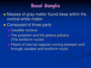
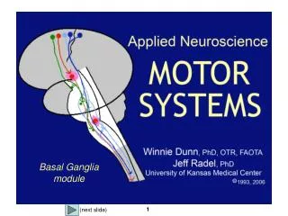
Basal Ganglia Definition: a group of nuclei, located deep within the cerebral hemispheres. They are part of the extrapyramidal motor system, principally involved in the control of posture and movements (primarily by inhibiting motor functions)
Components • The basal ganglia include: • 1-Caudate nucleus • 2-Putamen • 3-Globus pallidus: - external segment - internal segment • SubthalamicNucleus • Substantianigra Are functionally related to the basal ganglia • The Amygdala, located within the temporal lobe has a similar embryologic origin but functionally is part of the limbic system.
Basal Ganglia: Organization • Functionally the putamenis more closely allied to caudatenucleus and together constitute the neostriatumor simply STRIATUM. • The globuspallidusis the oldest part of corpus striatum and is called paleostriatumor simply PALLIDUM. • Anatomically the putamenand globuspallidusare together called the lentiform nucleus. The striatum and the pallidum are collectively known as the corpus striatum.
Corpus Striatum • Lies lateral to the thalamus. • It is divided by internal capsule into caudate nucleus and lentiform nucleus. • Bands of grey matter pass from lentiform nucleus across the internal capsule to the caudate nucleus, giving the striated appearance hence, the name corpus striatum. • The most ventral part of the corpus striatum is called the nucleus accumbens, which has connections with the limbic system.
Caudate Nucleus • Large C-shaped or comma-shaped grey mass • It has a Head, a Body and a Tail. Head : ( Anterior) Large, & rounded • Forms the lateral wall of the anterior horn of lateral ventricle. • Completely separated from the putamen by the internal capsule except rostrally where it is continuous with the putamen through and beneath the anterior limb of internal capsule.
Caudate Nucleus • Body: Long & narrow continuous with head • lies in the floor of body of lateral ventricle. • Tail : Long, narrow & tapering, • descends posteriorly into the temporal lobe • lies in the roof of inferior horn of lateral ventricle.
Lentiform Nucleus • A three sided, wedge-shaped mass of grey matter, with a convex outer surface and an apex which lies against the genuof the internal capsule • Divided into a: • Larger darker lateral portion called Putamen& • Smaller, lighter medial portion calledGlobuspallidus External capsule
Putamen * * • Lies lateral to the internal capsule and globuspallidus • Separated from globuspallidus by a thin sheath of Nerve Fibers, the lateral medullary lamina • The white matter lateral to putamen is divided, by a sheath of GREY MATTER, the CLAUSTRUM, into two parts: • The part between the putamen and claustrum forms the external capsule* • between the claustrum and the cortex of insula (deep within the lateral fissure) forms the extreme capsule*.
GlobusPallidus • Lies medial to the putamen, separated from it by lateral medullary lamina • Consists of two divisions, the lateral & the medial segments, separated by a thin sheath of nerve fibers, the medial medullary lamina. • The medial segment is similar, in terms of cytology and connections with the pars reticulataof substantianigra
Connections of the basal ganglia • Caudate nucleus • Putamen • Globus pallidus – output leaves receive input
Connections of striatum Caudate nucleus & putamen – INPUT Receive afferent - cerebral cortex, intralaminar thalamic nuclei, subs nigra Efferent – globuspallidus, subs nigra Connections of globuspallidus 2 segments – med & lat Med & subs nigra– OUTPUT Receive afferent – striatum, subthalamic nucleus Efferent Lat – subthalamic N Med – thalamus (VA, VM ,CM) – motor areas Connections of the basal ganglia
Connections of the Striatum Afferent Fibers Corticostriatal Thalamostriatal Nigrostriatal Brainstem striatal 1 2 I Efferent Fibers Striatopallidal Striatonigral 4 II 3
Connections of the GlobusPallidus Together with the pars reticulata of substatianigra, the medial segment is regarded as output part of the basal ganglia. Afferent Connections: • Striatopallidal fibers • Subthalamopallidal fibers: Originate from subthalamic nucleus of the diencephalon. Pass laterally through the internal capsule as subthalamic fasciculus and terminate in both segments of globuspallidus (more in the medial segment). 1 2 subthalamic fasciculus
Connections of the Globus Pallidus Efferent connections: • The two segments have different projections • The lateral segment principally projects to subthalamic nucleus via the subthalamic fascicle. • The medial segment together with the pars reticulataof substatianigra projects: • primarily to the thalamus (pallidothalamic fibers) • to the pedunculo-pontine nucleus of the brain stem tegmentum (pallidotegmental fibers) 1 2 Midbrain subthalamic fasciculus
Pallidothalamic fibers: • take two routes: • 1-Pass around the anterior margin of the internal capsule as the ansalenticularis • 2-Pass through the internal capsule as the lenticular fasciculus. • Both sets of fibers continue to course medially and then loop dorsally and laterally as thalamic fasciculus to enter the thalamus (VA, VL and centromedian nuclei).
All of this circuitry is on the same side of the brain—uncrossed. • Thus, the basal ganglia affect function mediated by the ipsilateral motor cortex. • Since motor cortex controls the movements of the contralateral body, the basal ganglia affects movements of the contralateral side of the body.
Connections i/laminar thal CN/
Functions of the basal ganglia • Cerebral cortex, basal ganglia, cerebellum and thalamus • motor activity • muscle tone • organization of movement • What type ? : cerebral cortex • How to perform? : basal ganglia + cerebellum • Assist in regulation : thalamus
Functions of the basal ganglia Function • Part of extra-pyramidal motor system • Facilitate behaviour & movement – required and appropriate • Inhibit unwanted & inappropriate • The deficits tend to fall into one of two categories: 1- The presence of extraneous unwanted movements • OR 2- An absence or difficulty with intended movements. The balance between the cerebellum and the basal ganglia allows smooth, coordinated movement, and a disturbance in either system will show up as movement disorders.
Cerebral cortex Corticospinal Corticobulbar Corticostriatal excitatory Direct Indirect inhibitory Striatonigral Striatopallidal Latpallidal inhibitory Subthalamic N inhibitory Med pallidal Activate neurone Disinhibit neurone thalamus Facilitate movement Inhibit unwanted movement
Diseases of basal ganglia • Change in muscle tone • Abnormal involuntary movement • Parkinsonism • Effect on the opposite side • Degeneration of dopamine-producing cells in substantianigra-depletion of dopamine in striatum • Resting tremors • Rigidity – simultaneous contraction of flexors and extensors • Bradykinesia = Slowness of movement – brake cannot be released • No paralysis, sensory loss, ataxia
Cerebral cortex Corticospinal Corticobulbar Corticostriatal excitatory Direct Indirect inhibitory Striatonigral Striatopallidal Lat pallidal inhibitory Subthalamic N inhibitory Med pallidal Activate neurone Disinhibit neurone thalamus Facilitate movement Inhibit unwanted movement
Basal Ganglia-SummaryFunction - Dysfunction • Its dysfunction Does NOT cause paralysis, sensory loss or ataxia, but leads to: • Abnormal motor control: emergence of abnormal, involuntary movements (dyskinesias) e.g. tremors, chorea, athetosis, myoclonus, tic or dystonia.. • Alteration in muscle tone (hypertonia /hypotonia). • Function • The corpus striatum assists in regulation of voluntary movement and learning of motor skills. • Their function is to facilitate behavior and movement that are required and appropriate, and inhibit unwanted or inappropriate movement.