Download
1 / 29
310 likes | 719 Views
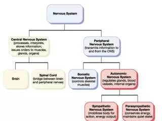
The Autonomic Nervous System. Chapter 16. Introduction. Makes all routine adjustments in physiological systems. Consists of visceral motor (efferent) neurons Involves 2 neurons synapsing in an autonomic ganglion Preganglionic (neuron #1) Postganglionic (neuron #2).

E N D
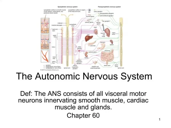
The Autonomic Nervous System Chapter 16
Introduction • Makes all routine adjustments in physiological systems. • Consists of visceral motor (efferent) neurons • Involves 2 neurons synapsing in an autonomic ganglion • Preganglionic (neuron #1) • Postganglionic (neuron #2)
Nerve Fibers of the ANS • Preganglionic (neuron #1) • Always myelinated • Neurotransmitter • ACh • Postganglionic (neuron #2) • Always nonmyelinated • Neurotransmitter • Ach • Norepinephrine
Subdivisions of the ANS • Sympathetic division (thoracolumbar) • Cell bodies for neurons #1 • thoracic and lumbar portions of the spinal cord. • T1 – L2 • Parasympathetic division (craniosacral) • Cell bodies • brain stem (cranial nerves) • sacral portion of the spinal cord.
Functions of the ANS • Sympathetic division • Stimulates heart beat & tissue metabolism, increases alertness, prepares the body to deal with emergencies (“fight or flight” division) • Parasympathetic division • Slows the heart rate, inhibits senses, prepares the body for rest and relaxation; (“rest and digest” division).
Organization of the SNS • Neurons #1 • Short • Usually synapse with neurons #2 (long) in ganglia • Ganglia • Sympathetic chain • Collateral
Sympathetic Chain Ganglia • A chain of ganglia that run alongside the spinal cord • Extends on both sides of the vertebral column • Carries preganglionic fibers and cell bodies of postganglionic neurons
Anatomy of the Sympathetic Chain • Rami communicates from spinal nerves connect to the chain • Splanchnic nerves project from the chain
Routes of Preganglionic Axons • Cell bodies of neurons #1 • In lateral gray horns • Axons of neurons #1 enter ventral root • Axons pass to spinal nerve • Axons leave spinal nerve via white and gray branches (rami communicates)
Routes of Preganglionic Axons • There are 4 possible routes that sympathetic neurons #1 may follow
Routes of Pregangleonic Axons – Possibility #1 • Neuron #1 synapses with the ganglion at same level of spinal cord • Neuron #2 leaves at that level via the gray ramus communicans, rejoins the same level spinal nerve
Routes of Preganglionic Axons - Possibility #2 • Neuron #1 goes up or down the chain and synapses at some other level. • Neuron #2 leaves at that level via the gray ramus communicans, rejoins the spinal nerve at that level.
Routes of Preganglionic Axons - Possibility #3 • Neuron #1 does not synapse in the chain but exits by a splanchnic nerve and synapses in a collateral ganglion. • Neuron #2 travels from that ganglion to its destination.
Collateral Ganglia • Location – anterior to the aorta in the abdominopelvic cavity • Celiac ganglion • Innervates upper abdominal viscera • Superior mesenteric ganglion • Innervates middle abdominal viscera • Inferior mesenteric ganglion • Innervates lower abdominal & pelvic organs
Routes of Preganglionic Axons – Possibility #4 • Neuron #1 does not synapse in a ganglion • Synapses with adrenal medulla • Only preganglionic neurons are in this pathway • Neuron #1 stimulates the medulla • The medulla releases norepinephrine and epinephrine (adrenaline) to blood
Effects of Sympathetic Stimulation • Widespread • The sympathetic chain allows one preganglionic fiber to synapse with many postganglionic neurons • Enhanced & prolonged by the adrenal medulla
Neurotransmitters • Preganglionic fibers release acetylcholine (Ach) • Cholinergic • Postganglionic fibers (most) release norepinephrine (NE) • Adrenergic • Adrenal medulla releases norepinephrine and epinephrine (adrenaline)
Membrane Receptors & Sympathetic Function • 2 types of receptors in synapses • The same neurotransmitter can have different effects • Alpha receptors cause a rise in intracellular calcium • Beta receptors cause changes in the metabolic activity of the target cells
Summary of Sympathetic Division • Cell bodies are found in the thoracic and lumbar portions of the spinal cord • Preganglionic fibers are short, connect to the sympathetic chain, and synapse with long postganglionic fibers • Preganglionic fibers produce ACh, postganglionic fibers produce NE or Ach • “Fight or flight” division
Organization of the PNS • Cell bodies are in the brain or in the gray matter of the spinal cord (sacral region) • Neurons #1 exit the cranial region through cranial nerves 3, 7, 9, & 10 • Neurons #1 exit the spinal cord through the sacral spinal nerves
Organization of the PNS • Neurons #1 are long and synapse with neurons #2 (short) in ganglia • Ganglia are found on near the visceral effector
Effects of Parasympathetic Stimulation • The cranial nerve fibers involved are motor - control smooth muscle & glands in the upper body
General Functions of the PNS • Prepares the individual for rest and relaxation • “Rest & digest” division • Effects on various organs: • Decreases heart rate • Constricts bronchioles • Increases salivation • Increases motility of stomach • Increases motility of colon • Constricts pupils
Neurotransmitter • Both preganglionic and postganglionic fibers release acetylcholine • Causes localized and short-term effects
Summary of the Parasympathetic Division • Cell bodies are found in the brain and in the sacral region of the spinal cord • Preganglionic fibers are long and synapse with short postganglionic fibers on or near the target viscera • Both preganglionic and postganglionic fibers produce Ach • “Rest & digest” division
Relationship Between the Sympathetic and Parasympathetic Divisions • Most organs receive dual innervation • Visceral organs are intrinsically excited • ANS either increase excitation or inhibit the activity • Eg. Heart rate