Download
1 / 39
400 likes | 638 Views
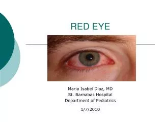
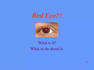
Red Eye . Grace Wong GPST1. Aims . Assessment of the red eye Common causes of red eye Painful and Non Painful Signs and symptoms Management of each condition. Red Eye. Common presentation in primary care and in A+E Most cases due to relatively trivial problems

E N D
Red Eye Grace Wong GPST1
Aims • Assessment of the red eye • Common causes of red eye • Painful and Non Painful • Signs and symptoms • Management of each condition
Red Eye • Common presentation in primary care and in A+E • Most cases due to relatively trivial problems • Most common is conjunctivitis • Small proportion are serious and need urgeny treatment • Sometimes difficulty in discerning between causes • Most practical way is; • Pain or not • Visual acuity
History • Onset • Pain • Visual Changes • Photophobia • Foreign body sensation • Trauma • Discharge, clear or colored • Bilateral or unilateral
Social history • Nursery school teacher • Co-morbid condition • Collagen vascular disorders • Rheumatoid, gout • TB, sarcoidosis • HTN • Past Ocular History • E.g. Similar episodes • Surgery • Lazy eye • Contact lenses
Examination • Visual acuity • Extra ocular movements • Pen light examination (reactivity, corneal opacity, pupil shape, discharge, infection) • Test for direct and consensual photophobia • Slit lamp examination – with and without fluorescein • Anterior chamber evaluation – depth, cells • IOP meaurements
Differential Diagnosis • Think systemically about the structures within the eye to common to differential diagnosis • Inflammation of orbit? • Lid Disease • Scleral inflammation • Corneal disease • Uveal/iris inflammation • Other e.g. glaucoma
Conjunctivitis • Most common cause of red eye • Inflammation of the conjunctiva • Sore red eye (gritty or itchy discomfort) • Discharge (clear, mucoid or muco-purulent) • Sticky eyelids • No visual changes • Unilateral or bilateral • Examination - enlarged papillae under upper eye lid or pre auricular lymph nodes
Causes • Allergic, viral or bacterial • Difficult to distinguish between types • Both bacterial and viral can occur after a viral URTI
Management - Infective • 85% of cases clear in <7 days with or without tx • Advise patients to bathe the affected eye with boiled cooled water am and pm • If symptoms not improve in >5 days • Swab for MC+S • Treat empirically with chloramphenicol QDS • consider alternative diagnosis e.g. allergy, dry eyes, Consider referral >7-10 days or if suspicion of herpetic infection
Management - Allergic • Topic or systemic anti histamines e.g. sodium cromoglicate eye drops • Avoid topical steroids – long term complications e.g. cataract, glaucoma, fungal infection • Consider cold compress and wash out with cold water during acute exacerbation Refer if symptoms are persistent despite treatment or if vision is affected
Subconjunctival Haemorrhage • Spontaneous painless localised haemorrhage under the conjunctiva • Common in the elderly • Spontaneous or traumatic • Looks alarming but generally painless (may cause some aching) • Clear spontaneously in 1-2 weeks but may recur
Associations • Hypertension • Clotting disorders • Leukaemia • Increased venous pressure • Check BP • If severe/recurrent • Check FBC and clotting screen
Signs • Blood under conjunctiva covering part or all of eye • Normal Visual Acuity • Consider referral if; • Follows trauma • More than a slight discomfort • Fails to settle spontaneously over 1 week
Blepharitis • Chronic low grade inflammation of meibomian glands and lid margins • Both eyes usually affected • Often associated with Dry eye syndrome, seborrhoeic dermatitis, rosacea
Causes • Staphylococcal • Seborrhoeic – associated with seborrhoeic dermatitis. Yeast is involved and can trigger inflammatory reaction • Meibomain – gland dysfunction unable to lubricate eye
Symptoms • Presents with long history of irritable burning dry red eyes • Eyelids have red margins • Look inflamed and greasy • Tiny flakes or scales on eyelids • Sticky with discharge • Meibomian glands may block an fill with oily fluid • Symptoms come and go
Treatment • Regular eyelid hygiene – warm, massage and cleansing • Remove scales and crusts from lid margins • Treat dry eye symptoms with preservative free tear supplements e.g. liquifilm • Antibiotic eye treatment if eyelid becomes infection e.g. fusidic acid (topical on eyelid). Can be up to 3 month course
Keratitis • Inflammation of the cornea • Bacterial, viral or fungal infections • Can be non infective e.g. trauma or auto-immune, dry eyes, entropion • History of contact lens wear • Previous episodes e.g. HSV infection
Signs • Very painful red eye • Photophobia • Foreign body sensation • Reduced visual acuity depends on nature of problem • Circumcornealinjection • Conjunctiva is also inflamed – keratoconjuncivitis • Discharge – water, mucoid or purulent • Pupil may be small • Fluorescinreadily demonstrates any ulceration
Complications • Significant loss of vision secondary to scarring or astigmastism • Complications can lead to blindness; • Corneal perforation • Choroidal detachment • Endopthalmitis • CORNEAL ULCERATION IS AN OPTHALMOLOGIC EMERGENCY
Treatment • The cause must be identified prior to treatment - some therapies benefit whilst others can harm • Refer the same day for urgent ophthalmological review • Delay may result in loss of sight
Caution • If caused by Herpes simplex infection and dendritic ulcer • AVOID topical steroids as can cause massive amoebic ulceration and blindness • Typical dendritic ulcer – delicate branching pattern
Scleritis • Severe inflammation that occurs throughout the entire thickness of the sclera • Rare • Average age 52 yrs • Can be unilateral or bilateral • Affects more women than men • Can affect anterior or posterior segment • Either nodular, diffuse or necrotizing
Associated • The sclera is an avascular structure • 50% is associated with systemic illness; • Herpes Zoster • Rheumatoid arthritis • SLE • Polyarteritisnodosum • Wegner’s granulomatosis • Trauma • Infection • Surgery
Presentation • Red eye • Severe boring eye pain – may radiate to forehead, brow or jaw • Key symptom; gradual onset (days or weeks) • Pain worse with movement of eye and at night • Watering • Photophobia • Decreased visual acuity • Eye is tender to touch and may have deep purple hue • There may be accompanying uveitis and keratitis
Treatment • Urgent referral to ophthalmology • Treated with steroids • Complications include • Cataract • Glaucoma • Retinal detachment
Iritis (anterior uveitis) • Most common in young/middle aged adults • Acute onset of pain • Increasing pain as eye converges and pupil constrict • Photophobia • Blurred vision • Decreased visual acuity • Watering • Circumcornealrednress • Small or irregular pupil • +hypopyon (pus causing white fluid level line)
Causes • Secondary to corneal graft rejection • Eye infections e.g. toxoplasmosis, herpes virus keratitis • 30% are associated with seronegativearthropathies e.g. AS
Management • Refer urgently to ophthalmology • Complications include; • Posterior synechiae (irregular pupil shape) • Glaucoma • Cataract Relapses are common
Red Flag signs of a Red Eye • Decreased visual acuity • Pain deep in the eye – not surface irritation • Photophobia • Absent or sluggish pupil response • Corneal Damage on fluorseceinstaining or opacification • History of trauma These need same day referral
Questions? • http://www.patient.co.uk/doctor/The-Red-Eye.htm