Download
1 / 38
590 likes | 1.71k Views
Red Eye. ASMPH LEC Group 6 Abad and Imperial Ophthalmology Clerkship Rotation: TMC. Outline. Pathophysiology Evaluation Common Causes of Red Eye Subconjunctival Hemorrhage Blepharitis Conjunctivitis Pterygium Phylctenulosis Episcleratis Keratitis Corneal Abrasion

E N D
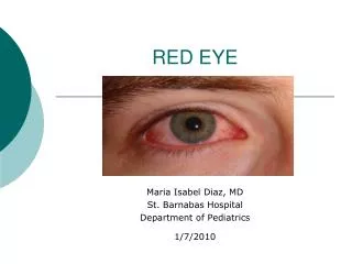
Red Eye ASMPH LEC Group 6 Abad and Imperial Ophthalmology Clerkship Rotation: TMC
Outline • Pathophysiology • Evaluation • Common Causes of Red Eye • Subconjunctival Hemorrhage • Blepharitis • Conjunctivitis • Pterygium • Phylctenulosis • Episcleratis • Keratitis • Corneal Abrasion • Acute Angle Glaucoma • Uveitis • Reference
Pathophysiology • Dilatation of blood vessels in the eye • conjunctival (superficial) • ciliary (deeper)
Evaluation • Chief complaint: RED EYE • HPI • Past Ocular History • Past Medical History • Ocular Exam
Subconjunctival hemorrhage Causes:Idiopathic, Trauma, Valsalva, Bleeding disorders, Drugs: Blood-thinners, steroids, contraceptives, Severe febrile systemic disease: Dengue, typhoid, malaria, etc. Usually benign and self- limiting Usually without pain and discharge; unilateral
Conjunctivitis • inflammation of the conjunctiva • dilatation of the superficial conjunctival blood vessels • hyperemia and edema with discharge
Adenoviral Conjunctivitis Usually self- limiting The common sore eye
Epidemic keratoconjunctivitis Common sequelae of adenoviral conjunctivitis. Serotypes 8, 11, 19 most common Treatment: artificial tears, cold compress, topical corticosteroids (controversial)
Gonococcal keratoconjunctivitis Neisseria gonorrhoeae: Hyper-acute, purulent conjunctivitis Rapid progression, copious purulent discharge, chemosis, lid edema Systemic IV/IM ceftriaxone (Cephalosporin) Topical antibiotics
Chlamydial (Inclusion) keratoconjunctivitis Chlamydia oculogenitalis Most common form of neonatal conjunctivitis and adult STD conjunctivitis Treatment: Oral doxycycline, topical erythromycin
Allergic conjunctivitis Hallmark: Itching! Type I hypersensitivity reaction (IgE-mediated) Treatment: Topical antihistamines, mast cell stabilizers and avoidance of allergen
Vernal conjunctivitis Common profile: Male, brown skin, under 20, lives at equatorial region. accumulation of eosinophil On palpebral conjunctiva, especially upper conjunctiva; Diffuse papillary hypertrophy: Giant (cobblestone) papillae Treatment: Topical antihistamines, mast cell stabilizers, corticosteroids FOR SHORT TERM; self-limiting
Giant Papillary Conjunctivitis Usually occurs in soft contact lens wearers: Contact lens material, solution, debris Treatment: Discontinuation of contact lens, topical antihistamine, mast cell stabilizers, shift to disposable lenses.
Pterygium The redness is confined largely to a raised, yellowish, fleshy lesion that is usually located on the nasal side of the bulbar conjunctiva Benign fibrovascular proliferation covered by conjunctival-like epithelium extending into peripheral cornea Location: Within or Above Bowman’s Line Treatment: Surgery, Excision with ancillary procedure
Phylctenulosis Symptoms: tearing, ocular irritation, mild to severe photophobia and a history of similar episodes Focal, translucent lymphocytic nodules generally located at limbus Cause: Delayed Cell-Mediated Hypersensitivity (IV) Treatment: Improve Eyelid Hygiene, Topical Corticosteroids
Episcleritis Simple: intermittent bouts of moderate-to-severe inflammation that often recur at 1- to 3-month intervals Nodular: prolonged attacks of inflammation that are typically more painful than simple episcleritis Inflammatory condition affecting the episcleral tissue Symptoms: Rapid onset of redness, dull ache, and tenderness on palpation Treatment: Topical Vasoconstrictors, Mild Corticosteroids
Bacterial Keratitis • Inflammation of the cornea due to infection • Symptoms • Pain and foreign body sensation due to mechanical effects of lids • Watering from the eye due to reflex hyperlacrimation • Photophobia from stimulation of nerve endings • Blurred vision from corneal haze • Redness of eyes due to congestion of circumcorneal vessels
Bacterial KeratitisStreptococcus pneumoniae Very painful! Serpiginous, gray-white stromal infiltrate and hypopyon characteristic of Gram-positive bacteria Suppuration does not usually extend over entire corneal surface Treatment: Topical erythromycin, chloramphenicol, 4th generation fluoroquinolones (moxiflocxcin, gatifloxacin), Oral cephalosporin, erythromycin, Cypoplegics
Bacterial KeratitisPseudomonas aeruginosa Common in immunocompromised patients, contact lens wearers with faulty hygiene Typical Gram-negative corneal ulcer: Rapid evolution, marked tendency to spread. Can perforate in 48 hours. Treatment: Topical tobramycin, ciprofloxacin, moxifloxacin, gatifloxacin
Fungal Keratitis • Intense suppuration, progressive hypopyon • Modes of infection: • Injury by vegetative material such as crop, leaf, branch of tree, straw, hay or decaying vegetable matter. • Common sufferers are field workers especially during harvest season • Therapeutic problem: No effective topical agent • Debridement: Scrape it off and reduce load of organism or perform keratectomy. • Candida: Natamycin; ketoconazole, voriconazole, amphotericin B
Fungal Keratitis Yeast Fungi Filamentous Fungi
Herpes simplex keratitis Coalesces in a few days into branching or dendritic lesion • Mode of infection: • HSV1 - Through kissing or coming in close contact with patient suffering from herpes labialis. • HSV2 - Transmitted to eyes of neonates through infected genitalia of the mother. Symptoms: Injection, Irritation, Mucoid discharge, Pain, Mild photophobia Treatment: Self limited but recurrent. Topical/systemic acyclovir, ganciclovir, debridement
Corneal abrasion Symptoms: Acute pain after ocular trauma Photophobia, excessive tearing, blepharospasm, foreign body sensation, blurred vision Follows Occular Trauma May be superficial or deep Treatment: Patching, Topical Antibiotics, Cycloplegics
Acute Angle Closure Glaucoma Symptoms Signs • ocular pain, headache • unilateral blurring of vision • "iridescent" vision: haloes around lights • nausea and vomiting • Elevated intraocular pressure (>40 mmHg) • deep circumlimbalconjunctival and episcleral injection: "ciliary flush" • fixed, mid-dilated pupil • edematous or steamy cornea • shallow anterior chamber
Acute Angle Closure Glaucoma • Treatment: Lower IOP • Carbonic anhydrase inhibitors • Hyperosmotic agents • Pilocarpine • Supportive: steroids and analgesics • Laser Iridotomy
Acute anterior uveitis Hallmark: Cells and Flare • Signs • Ciliary flush • Sterile hypopyon (severe) • Cells and flares • Keratic precipitates • Posterior synechiae • Granulomatous nodules • Symptoms • Deep, dull pain of • involved eye and surrounding orbit • Photophobia • Tearing • Difficulty in reading Uveitis: Inflammation of one or all parts of the uveal tract
Acute anterior uveitis Systemic causes Infectious causes • Ankylosingspondylitis • Bechet’s disease • Chronic granulomatous disease • Enthisitis • Inflammatory bowel disease • Kawasaki’s disease • Multiple sclerosis • Polyarteritisnodosa • Psoriatic arthritis • SLE • Vogt-Koyanagi-Harada syndrome • Brucellosis • Herpes simplex • Herpes zoster • Leptospirosis • Lyme disease • Syphilis • Toxoplasmosis • Tuberculosis
Acute anterior uveitis • Treatment • Immobilize iris, ciliary body to relieve pain (ie. atropine, cyclopentolate) • Reduce inflammation (ie. topical steroids) • Treat underlying ocular, systemic disease
References • Vaughan & Asbury’s General Ophthalmology 17th ed. • ASMPH Ophthalmology Lecture Notes on “Common Causes of Red Eye” by Dr. Victor L. Caparas. January 2010. • The Red Eye. The New England Journal of Medicine. Volume 343 Number 5. December 2007.