Download
1 / 29
300 likes | 352 Views
Learn about carcinoma of the colon, its types, staging, clinical features, spread, investigations, treatment options, and relevant pathology. Understand the staging criteria, including Dukes classification and TNM staging. Discover the various surgical procedures like hemicolectomy and chemotherapy options like FOLFOX and FOLFIRI.

E N D
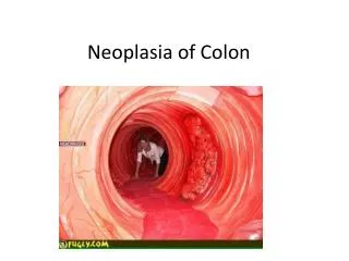
Carcinoma of colon Etiology: Diet- high protein , high fat diet, Fish, Beer, Low fibre diet : Intestinal bacteria :Genetic - siblings, children.FAP, HNPCC, PeutzJegher’s syndrome :Inflammatory bowel diseases :Radiation : Depressed immunity :Ureterocolicanastomosis
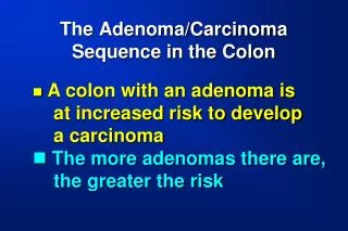
Normal colonic mucosa>dysplastic aberrant crypt foci>early adenoma>intermediate adenoma>late adenoma>ca in situ>invasive ca • Tumour suppressor genes- APC gene, p53 gene, STK 11 gene. • Mismatch Repair Gene, microsatellite instability. • Oncogenes- k ras.
Pathology • Adenocarcinoma(majority), undifferentiated carcinoma, neuroendocrine tumours, spindle cell tumour, squamous cell carcinoma, adenosquamous carcinoma • Common in distal colon: Sigmoid 21%, descending colon 5%, transverse colon12%, Ascending & caecum 25%, Each flexures 2% • Synchronous 3% and Metachronous 5%
Gross: Polypoidal/ cauliflower, Ulcerative, Annular/stenosing, Diffuse infiltrating
Microscopy: arise from the columnar epithelium or crypts of Liberkunh. Glandular formation. Varying degrees of differentiation (well, moderately, poorly).
Spread • Direct- along mucosa and submucosa, surrounding bowel, bladder, peritoneum, anterior abdominal wall • Lymphatic- epicolic nodes, paracolic, intermediate, main lymph nodes, para aortic • Haematogenous- liver, lungs, adrenals, kidneys, bones • Transcoelomic spread
Staging • Dukes: A- tumour confined to bowel wall B- spread beyond serosa to adjacent structures, no nodes C- Lymph nodes involved • TNM:Tx- primary cannot be assessed, T0- no evidence of primary, T1- tumour invades uptosubmucosa, T2- tumouruptomuscularispropria, T3- invades uptosubserosa or uptopericolic or pararectal tissue at unperitonealised area, T4- invasion of adjacent organ or perforation
N0- no regional nodes, N1- 1 to 3 nodes, N2- metastasis to 4 or more regional nodes • M0 , M1
Clinical Features • > 50 years • More in men • Depends on the site of tumour
Caecum & Asc Colon • Asymptomatic • Anorexia, Anemia, Asthenia • Rt Iliac Fossa pain • Increasing constipation • Nausea, Vomiting • Blood and mucous in stools • Intestinal obstruction • Mass in RIF • Acute appendicitis • Intussusception
Transverse colon • Vague symptoms • Constipation • Diarrhoea, Borborygmi • Intestinal obstruction
Descending colon • Increasing constipation • Alternating constipation with diarrhoea- blood and mucous • Left sided abdominal pain • Mass abdomen • Abdominal distension
Sigmoid colon • Progressive constipation • Pain abdomen • Tenesmus • Bleeding PR • Spurious diarrhoea • Sciatica • Perforation- peritonitis, paracolic abscess • Colo- vesical/ enteric/ cutaneous fistula
Investigations • F O B • USG abdomen • Sigmoidoscopy/Colonoscopy, biopsy • Ba Enema • C T scan • I V P • C E A
Treatment • Wide Resection or palliative resection/bypass • Preparation for surgery: Mechanical cleansing, Sterilisation of bowel (antibiotics), Hydration and electrolyte correction
Operations • Right Hemicolectomy- tumours of Caecum, ascending colon • Extended right hemicolectomy- Hepatic flexure, transverse colon, splenic flexure • Left hemicolectomy- splenic flexure, descending colon, sigmoid • Anterior resection- sigmoid colon. • Hepatic resections • Colostomy- transverse/ sigmoid • Ileo transverse anastomosis
Intestinal obstruction or perforations Lt side growth: primary resection, EEA , proximal colostomy/ Resection, end colostomy and mucous fistula/ Hartmann’s operation. Rt sided growth: Rthemicolectomy and primary anastomosis
Chemotherapy- FOLFOX( 5 FU, Leucovorin, Oxaliplatin), FOLFIRI(5FU, Leucovorin, Irinotecan). Targeted therapy with bevacizumab (VEGF inhibitor) or cetuximab (EGFR inhibitor) in patients with k ras mutation. • Radiotherapy- no significant role.