Download
1 / 21
220 likes | 256 Views
Explore the structure of cell membranes, the role of phospholipids and membrane proteins, and the evolution of membrane models. Learn about the fluid mosaic model, phospholipid bilayer, and membrane proteins.

E N D
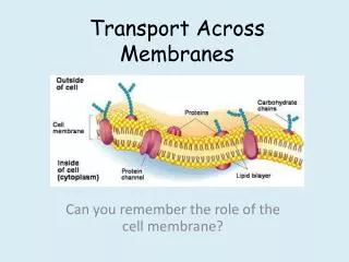
Transport across membranes Biology Mr Atkinson
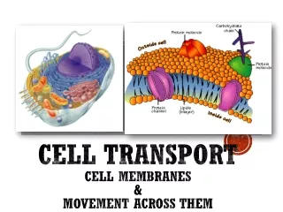
Objectives • Draw and Label a diagram to show the structure of membranes • Explain how the hydrophobic and hydrophilic properties of phospholipids helps to maintain the structure of cell membranes. • List the functions of membrane proteins.
Evidence for the Davson–Danielli model When clear electron micrographs of membranes became available, they appeared to show support for Davson–Danielli’s model, showing a three-layered structure. intracellular space (blue) This was taken to be the phospholipid bilayer (light) surrounded by two layers of protein (dark). 1st cell membrane 1 light layer = phospholipid bilayer 2 dark layers: protein 2nd cell membrane
Problems with the Davson–Danielli model By the end of the 1960s, new evidence cast doubts on the viability of the Davson–Danielli model. • The amount and type of membrane proteins vary greatly between different cells. • It was unclear how the proteins in the model would permit the membrane to change shape without bonds being broken. • Membrane proteins are largely hydrophobic and therefore should not be found where the model positioned them: in the aqueous cytoplasm and extracellular environment.
Evidence from freeze-fracturing In 1966, biologist Daniel Branton used freeze-fracturing to split cell membranes between the two lipid layers, revealing a 3D view of the surface texture. E-face: looking up at outer layer of membrane This revealed a smooth surface with small bumps sticking out. These were later identified as proteins. P-face: looking down on inner layer of membrane
The fluid mosaic model E-face The freeze-fracture images of cell membranes were further evidence against the Davson–Danielli model. They led to the development of the fluid mosaic model, proposed by Jonathan Singer and Garth Nicholson in 1972. P-face protein This model suggested that proteins are found within, not outside, the phospholipid bilayer.
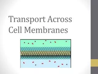
It is said to have a ‘fluid-mosaic’ structure • It is made of a phospholipid bilayer • With proteins floating among them • Some of the proteins have carbohydrates attached to them
Phospholipids: Structure and Function • Hydrophilic head • Hydrophobic Tail • Forms a bilayer
Structural Properties of Phospholipid Bilayer • Phospholipids are held together in a bilayer by hydrophobic interactions (weak associations) • Hydrophilic / hydrophobic layers restrict entry and exit of substances • Phospholipids allow for membrane fluidity / flexibility (important for functionality)
More structural properties • Phospholipids with short or unsaturated fatty acids are more fluid • Phospholipids can move horizontally or occasionally laterally to increase fluidity • Fluidity allows for the breaking / remaking of membranes (exocytosis / endocytosis)
Cholesterol Help with membrane fluidity Allow for effective functioning at a range of temperatures Plant cells do not have cholesterol, they rely on fatty acids to maintain fluidity.
Proteins • Two types of major proteins: • Integral proteins • Peripheral proteins
Functions of proteins • TRACIE • T – Transport • R - Receptors • A – Anchorage • C – Cell recognition • I – Intercellular Joining • E – Enzymatic activity
Objectives • Draw and Label a diagram to show the structure of membranes • Explain how the hydrophobic and hydrophilic properties of phospholipids helps to maintain the structure of cell membranes. • List the functions of membrane proteins.
Further reading • IB Bio ninja: http://ib.bioninja.com.au/standard-level/topic-2-cells/24-membranes.html • Click4biology: http://click4biology.info/c4b/2/cell2.4.htm • IB Guides: • http://ibguides.com/biology/notes/membranes