Download
1 / 47
530 likes | 1.12k Views
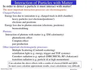
Interaction of X-rays with Matter and Imaging. Gocha Khelashvili Assistant Research Professor of Physics Illinois Institute of Technology Research Physicist EXELAR Medical Corporation. The Plan. X-ray Interactions with Matter Used at Imaging Energies

E N D
Interaction of X-rays with Matter and Imaging Gocha Khelashvili Assistant Research Professor of Physics Illinois Institute of Technology Research Physicist EXELAR Medical Corporation
The Plan • X-ray Interactions with Matter Used at Imaging Energies Photoelectric Effect Coherent Scattering Incoherent Scattering Refraction Small- and Ultra-small Angle Scattering • Radiography How does it work? Imaging Parameters and Sources of X-ray contrast Drawbacks of Radiography • Diffraction Enhanced Imaging (DEI) How does it work? Imaging Parameters and Sources of X-ray contrast Drawbacks of DEI • Multiple Image Radiography (MIR-Planar Mode) How does it work? Sources of X-ray contrast MIR parameters and images • MIR Model Based on Discrete Scatterers Multiple scattering series approach and MIR transport equation Solution of MIR transport equation Imaging Parameters • Laboratory DEI / MIR Machine • Summary
Effects of Binding Energy in Compton (Incoherent) Scattering
Effects of Binding Energy in Compton (Incoherent) Scattering
Radiography Setup and Imaging Principles Radiology Setup Object Double Crystal Monochromator Si(333) Incident X-ray beam Area Detector Attenuation Law Image Image Contrast
Drawbacks of Radiography Incoherently Scattered Beam Detector Pixel Object Pixel Attenuated Beam (by absorption) Image Contrast
DEI Setup and Imaging Principles DEI Setup Area Detector Object Double Crystal Monochromator Si(333) Analyzer Crystal Si(333) Incident X-ray beam
Formation of DE Images Incoherently Scattered Beam is Blocked by Crystal Detector Pixel Object Pixel Enhanced Attenuated Beam
Physics of DEI Pisano, Johnston(UNC); Sayers(NCSU); Zhong (BNL); Thomlinson (ESRF); Chapman(IIT) Low Angle Side High Angle Side 1.00 0.80 0.60 Relative Intensity I/Io 0.40 0.20 0.00 -10 -5 0 5 10 Analyzer Angle (mradians) Data from NSLS X27
Calculation of DEI Images Low Angle Side High Angle Side 1.00 0.80 0.60 Relative Intensity I/Io 0.40 0.20 0.00 -10 -5 0 5 10 Analyzer Angle (mrad)
6 1 0 - 0 5 4 DEI Comparison - Conventional and DEIACR - Phantom Map Conventional
DEI image of ACR phantom - smallest calcifications Data from NSLS X27
Cancer in Breast Tissue Pisano, Johnston(UNC); Sayers(NCSU); Zhong (BNL); Thomlinson (ESRF); Chapman(IIT) Conventional DEI - Absorption DEI - Refraction BNL Sept 1997
Drawbacks of DEI Detector Pixel Object Pixel
600 Rod, off-center 600 Background 400 400 200 200 -1 -0.6 -0.2 0.2 0.6 1 -5 x 10 -1 -0.6 -0.2 0.2 0.6 1 -5 x 10 600 Thick Paper 600 Rod and Paper 400 400 200 200 -1 -0.6 -0.2 0.2 0.6 1 -5 x 10 1 -1 -0.6 -0.2 0.2 0.6 -5 x 10 Experimental Results
Refraction images Profiles no paper 1 MIR 0.8 MIR 0.6 0.4 DEI DEI thin paper DEI 0.2 thick paper 0 0 50 100 150 200 Position (pixels)
Discrete Scatterer Model Khelashvili, Brankov (IIT), Chapman (U.Sask), Anastasio, Yang (IIT), Zang (BNL), Wernick (IIT) Object Voxel
Multiple Ultra-Small Angle Scattering • Radiation Transport Theory Approach
Phase Function Khelashvili, Brankov (IIT), Chapman (U.Sask), Anastasio, Yang (IIT), Zang (BNL), Wernick (IIT)
Plane Wave Solution Khelashvili, Brankov (IIT), Chapman (U.Sask), Anastasio, Yang (IIT), Zang (BNL), Wernick (IIT)
Plane Wave Solution Khelashvili, Brankov (IIT), Chapman (U.Sask), Anastasio, Yang (IIT), Zang (BNL), Wernick (IIT)
Imaging Parameters Khelashvili, Brankov (IIT), Chapman (U.Sask), Anastasio, Yang (IIT), Zang (BNL), Wernick (IIT)
Experimental Conformation Khelashvili, Brankov (IIT), Chapman (U.Sask), Anastasio, Yang (IIT), Zang (BNL), Wernick (IIT) Lucite container – wedge shaped. Polymethylmethacrylate (PMMA) microspheres in glycerin.
Experimental Conformation Khelashvili, Brankov (IIT), Chapman (U.Sask), Anastasio, Yang (IIT), Zang (BNL), Wernick (IIT)
X-ray Source Detector Analyzer Pre-mono & Mono labDEI System Morrison, Nesch, Torres, Khelashvili (IIT), Hasnah (U. Qatar) Chapman (U.Sask)
1cm cartilage bone Lab DEI System tissue images Morrison, Nesch, Torres, Khelashvili, Chapman (IIT) Muehleman(Rush Medical College)
Summary • First reliable Theoretical Model of DEI – MIR has been developed. • Model can be used to simulate experiments starting from source, through crystals (this was known), through object (was unknown), through analyzer crystal (partially known – dynamical theory of diffraction – but crystal and beam specific calculations need to be done). • CT reconstructions – some steps are already taken in this direction – Miles N. Wernick et al “Preliminary study of multiple-image computed tomography” • CSRRI (IIT) / Nesch LLC – are developing in-lab research DEI instrument
Acknowledgements Funded by NIH/NIAMS. L.D. Chapman (Anatomy and Cell Biology, University of Saskatchewan, Canada) J. Brankov, M. Wernick, Y. Yang, M. Anastasio (Biomed. Engineering, IIT) T. Morrison and I. Nesch (CSRRI, IIT) C. Muehleman (Department of Anatomy and Cell Biology, Rush Medical College)