Download

1 / 51
510 likes | 528 Views
Learn the basics of interpreting Pulmonary Function Tests, including components, indications, and common patterns, to apply in patient care. Topics include spirometry, lung volumes, and diffusing capacity, with indications for diagnosis, screening, and monitoring. 8 Relevant

E N D
Pulmonary Function TestingThe Basics of Interpretation Jennifer Hale, M.D. Valley Baptist Family Practice Residency
Objectives • Identify the components of PFTs • Describe the indications • Develop a stepwise approach to interpretation • Recognize common patterns • Apply this information to patient care
Pulmonary Function TestingJennifer Hale, M.D. Which of the following is used to follow disease severity in COPD patients? • Total lung capacity (TLC) • Degree of responsiveness to bronchodilators • Forced vital capacity (FVC) • Forced expiratory volume in 1 second e. Diffusing capacity (DLCO)
Pulmonary Function TestingJennifer Hale, M.D. A 36yo WF, non-smoker, presents to your office for follow-up of ‘recurrent bronchitis.’ You suspect asthma and decide to order spirometry. Which of the following would you include in your prescription for testing? • Diffusing Capacity (DLCO) • If no obstruction present, add trial of bronchodilator • If no obstruction present, perform methacholine challenge • Flow volume loop • b and c
Pulmonary Function TestingJennifer Hale, M.D. A 68yo HM is admitted to the ICU with acute respiratory distress. A CXR obtained in the ED demonstrates bilateral pulmonary infiltrates, and his DLCO is elevated. What is the most likely diagnosis? • Pulmonary edema • Aspiration pneumonitis • Pulmonary emboli • Alveolar hemorrhage e. Interstitial lung disease
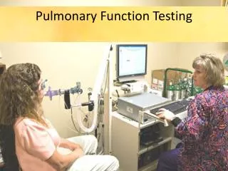
The Purpose Provide quantifiable, reproducible measurement of lung function
Description • Spirometry • Flow Volume Loop • Bronchodilator response • Lung volumes • Diffusion capacity (DLCO) • Bronchoprovocation testing • Maximum respiratory pressures • Simple and complex cardiopulmonary exercise testing
Indications — Diagnosis • Evaluation of signs and symptoms - SOB, exertional dyspnea, chronic cough • Screening at-risk populations • Monitoring pulmonary drug toxicity • Abnormal study - CXR, EKG, ABG, hemoglobin • Preoperative assessment
Indications — Diagnosis • Evaluation of signs and symptoms - SOB, exertional dyspnea, chronic cough • Screening at-risk populations • Monitoring pulmonary drug toxicity • Abnormal study - CXR, EKG, ABG, hemoglobin • Preoperative assessment Smokers > 45yo (former & current)
Indications — Diagnosis • Evaluation of signs and symptoms - SOB, exertional dyspnea, chronic cough • Screening at-risk populations • Evaluation of occupational symptoms • Monitoring pulmonary drug toxicity • Abnormal study - CXR, EKG, ABG, hemoglobin • Preoperative assessment
Indications — Prognostic • Assess severity • Follow response to therapy • Determine further treatment goals • Referral for surgery • Disability
Spirometry • Simple, office-based • Measures flow, volumes • Volume vs. Time • Can determine: - Forced expiratory volume in one second (FEV1) - Forced vital capacity (FVC) - FEV1/FVC - Forced expiratory flow 25%-75% (FEF25-75)
Obstructive Pattern • Decreased FEV1 • Decreased FVC • Decreased FEV1/FVC - <70% predicted • FEV1 used to follow severity in COPD
Obstructive Lung Disease — DifferentialDiagnosis • Asthma • COPD - chronic bronchitis - emphysema • Bronchiectasis • Bronchiolitis • Upper airway obstruction
Restrictive Pattern • Decreased FEV1 • Decreased FVC • FEV1/FVC normal or increased
Restrictive Lung Disease —DifferentialDiagnosis • Pleural • Parenchymal • Chest wall • Neuromuscular
Bronchodilator Response • Degree to which FEV1 improves with inhaled bronchodilator • Documents reversible airflow obstruction • Significant response if: - FEV1 increases by 12% and >200ml • Request if obstructive pattern on spirometry
Flow Volume Loop • “Spirogram” • Measures forced inspiratory and expiratory flow rate • Augments spirometry results • Indications: evaluation of upper airway obstruction (stridor, unexplained dyspnea)
Upper Airway Obstruction • Variable intrathoracic obstruction • Variable extrathoracic obstruction • Fixed obstruction
Lung Volumes • Measurement: - helium - nitrogen washout - body plethsmography • Indications: - Diagnose restrictive component - Differentiate chronic bronchitis from emphysema
Lung Volumes – Patterns • Obstructive - TLC > 120% predicted - RV > 120% predicted • Restrictive - TLC < 80% predicted - RV < 80% predicted
Diffusing Capacity • Diffusing capacity of lungs for CO • Measures ability of lungs to transport inhaled gas from alveoli to pulmonary capillaries • Depends on: - alveolar—capillary membrane - hemoglobin concentration - cardiac output
Decreased DLCO(<80% predicted) Obstructive lung disease Parenchymal disease Pulmonary vascular disease Anemia Increased DLCO (>120-140% predicted) Asthma (or normal) Pulmonary hemorrhage Polycythemia Left to right shunt Diffusing Capacity
DLCO — Indications • Differentiate asthma from emphysema • Evaluation and severity of restrictive lung disease • Early stages of pulmonary hypertension • Expensive!
Case1 CC/HPI:A 36yo WM, nonsmoker, presents to your clinic with c/o episodic cough for 6mo. Also reports occasional wheezing and dyspnea with exertion during softball practice. Exam: Heart RRR, no murmurs; Lungs CTAB, no labored breathing Based on your exam and a thorough review of systems, you suspect asthma and decide to order spirometry for further evaluation.
Continued… PFTs: FEV1 86% predicted FEV1/FVC 82% predicted Flow Volume Loop: normal inspiratory and expiratory pattern You still suspect asthma. What is your next step in the workup of this patient?
Bronchoprovocation • Useful for diagnosis of asthma in thesetting of normal pulmonary function tests • Common agents: - Methacholine, Histamine, others • Diagnostic if: ≥20% decrease in FEV1
Continued… SYMPTOMS ↓ PFTs ↓ OBSTRUCTION? ↓ ↓ YES NO ↓ ↓ BRONCHOPROVOCATION TREAT ↓ ↓ Obstruction? TREAT No Obstruction? Other Diagnosis
PFT Interpretation Strategy • What is the clinical question? • What is “normal”? • Did the test meet American Thoracic Society (ATS) criteria? • Don’t forget (or ignore) the flow volume loop!
Obstructive Pattern — Evaluation • Spirometry • FEV1, FVC: decreased • FEV1/FVC: decreased(<70% predicted) • FV Loop“scooped” • Lung Volumes • TLC, RV: increased • Bronchodilator responsiveness
Restrictive Pattern – Evaluation • Spirometry • FVC, FEV1: decreased • FEV1/FVC: normal or increased • FV Loop“witch’s hat” • DLCO decreased • Lung Volumes • TLC, RV: decreased • Muscle pressures may be important
Emphysema FEV1/FVC <70% “Scooped” FV curve TLC increased Increased compliance DLCO decreased Chronic Bronchitis FEV1/FVC <70% “Scooped” FV curve TLC normal Normal compliance DLCO usually normal PFT Patterns
PFT Patterns • Asthma • FEV1/FVC normal or decreased • DLCO normal or increased But PFTs may be normal bronchoprovocation
Pulmonary Function TestingJennifer Hale, M.D. Which of the following is used to follow disease severity in COPD patients? • Total lung capacity (TLC) • Degree of responsiveness to bronchodilators • Forced vital capacity (FVC) • Forced expiratory volume in 1 second e. Diffusing capacity (DLCO)
Pulmonary Function TestingJennifer Hale, M.D. Which of the following is used to follow disease severity in COPD patients? • Total lung capacity (TLC) • Degree of responsiveness to bronchodilators • Forced vital capacity (FVC) • Forced expiratory volume in 1 second e. Diffusing capacity (DLCO)
Pulmonary Function TestingJennifer Hale, M.D. A 36yo WF, non-smoker, presents to your office for follow-up of ‘recurrent bronchitis.’ You suspect asthma and decide to order spirometry. Which of the following would you include in your prescription for testing? • Diffusing Capacity (DLCO) • If no obstruction present, add trial of bronchodilator • If no obstruction present, perform methacholine challenge • Flow volume loop • b and c
Pulmonary Function TestingJennifer Hale, M.D. A 36yo WF, non-smoker, presents to your office for follow-up of ‘recurrent bronchitis.’ You suspect asthma and decide to order spirometry. Which of the following would you include in your prescription for testing? • Diffusing Capacity (DLCO) • If no obstruction present, add trial of bronchodilator • If no obstruction present, perform methacholine challenge • Flow volume loop • b and c
Pulmonary Function TestingJennifer Hale, M.D. A 68yo HM is admitted to the ICU with acute respiratory distress. A CXR obtained in the ED demonstrates bilateral pulmonary infiltrates, and his DLCO is elevated. What is the most likely diagnosis? • Pulmonary edema • Aspiration pneumonitis • Pulmonary emboli • Alveolar hemorrhage e. Interstitial lung disease
Pulmonary Function TestingJennifer Hale, M.D. A 68yo HM is admitted to the ICU with acute respiratory distress. A CXR obtained in the ED demonstrates bilateral pulmonary infiltrates, and his DLCO is elevated. What is the most likely diagnosis? • Pulmonary edema • Aspiration pneumonitis • Pulmonary emboli • Alveolar hemorrhage e. Interstitial lung disease