Download
1 / 105
1.08k likes | 1.32k Views
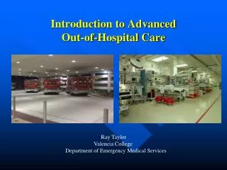
Division 1 Introduction to Advanced Prehospital Care. Chapter 4 General Principles of Pathophysiology Part I How Normal Body Processes Are Altered by Disease and Injury. Topics. Disease Risk Hypoperfusion Shock Multiple Organ Dysfunction Syndrome. Pathophysiology.

E N D
Chapter 4General Principles of PathophysiologyPart I How Normal Body Processes Are Altered by Disease and Injury
Topics • Disease Risk • Hypoperfusion • Shock • Multiple Organ Dysfunction Syndrome
Pathophysiology • The study of how diseases alter the normal physiological processes of the human body • From the root “patho” meaning disease
Cellular Adaptation • Cells, tissues, organs, and organ systems can adapt to both normal and injurious conditions. • Adaptation to external stressors results in alteration of structure and function. • Examples: Growth of the uterus during pregnancy, dilation of the left ventricle after an MI.
Types of Cellular Adaptations(1 of 2) • Atrophy • Decreased size resulting from a decreased workload • Hypertrophy • An increase in cell size resulting from an increased workload
Types of Cellular Adaptations(2 of 2) • Hyperplasia • An increase in the number of cells resulting from an increased workload • Metaplasia • Replacement of one type of cell by another type of cell that is not normal for that tissue • Dysplasia • A change in cell size, shape, or appearance caused by an external stressor
Cellular Injury • Hypoxic • Chemical • Infectious • Immunologic/Inflammatory • Physical agents • Nutritional balances • Genetic factors
Manifestation of Cellular Injury • When cells are injured metabolism is changed, causing substances to infiltrate or accumulate to an abnormal degree in cells.
Cellular Swelling • Results from a permeable or damaged cellular membrane • Caused by an inability to maintain stable intra- and extracellular fluid and electrolyte levels
Fatty Change • Lipids invade the area of injury. • Occurs most commonly in vascular organs, most frequently the liver. • Causes a disruption of the cellular membrane and metabolism and interferes with the vital functions of the organ.
Signs and Symptoms of Cellular Change • Fatigue and malaise • Altered appetite • Fever • Increased heart rate associated with fever • Pain
Cell Death (1 of 3) • Apoptosis • Injured cell releases enzymes that engulf and destroy the cell. • Cells shrink. • Eliminating damaged and dead cells allows tissues to repair and possibly regenerate.
Cell Death (2 of 3) • Necrosis • A pathological process • Cells swell and rupture • Coagulative • Liquefactive • Caseous • Fatty
Cell Death (3 of 3) • Gangrenous necrosis • Cell death over a wide area • Dry • Wet • Gas
Where the Water Is Found • Intracellular fluid—fluid inside the cells • Extracellular fluid—all the fluid outside the body cells • Intravascular fluid—fluid within the circulatory system • Interstitial fluid—fluid outside of the cell membranes but not within the circulatory system
Edema • Accumulation of water in the interstitial space due to disruption in the forces and mechanisms that normally keep net filtration at zero
Mechanisms That Cause Edema • A decrease in plasma oncotic force • An increase in hydrostatic pressure • Increased capillary permeability • Lymphatic channel obstruction
Edema (1 of 2) • Can be local or within a certain organ system • Sprained ankle vs. pulmonary edema
Edema (2 of 2) • Water in interstitial spaces is not available for metabolic processes. • Edema, therefore, can cause a relative condition of dehydration.
The percentage of the blood occupied by the red blood cells is termed the hematocrit.
Transfusion Reactions • Transfusion reactions occur when there is a discrepancy between the blood type of the patient and the type of the blood being transfused.
Fever Chills Hives Hypotension Palpitations Tachycardia Flushing of the skin Headache Loss ofconsciousness Nausea Vomiting Shortness of breath Signs and Symptoms of Transfusion Reactions
Treatment of Transfusion Reactions (1 of 2) • IMMEDIATELY stop the transfusion. • Save the substance being transfused. • Rapid IV infusion.
Treatment of Transfusion Reactions (2 of 2) • Assess the patient’s mental status. • Administer oxygen. • Contact medical direction. • Be prepared to administer mannitol, diphenhydramine, or furosemide.
Hemoglobin-Based Oxygen-Carrying Solutions (HBOCs) • Commonly referred to as “blood substitutes” • Compatible with all blood types • Do not require blood typing, testing, or cross-matching • PolyHeme • Hemopure
Colloids • Colloids remain in intravascular spaces for an extended period of time and have oncotic force. • Plasma protein fraction (Plasmanate) • Salt-poor albumin • Dextran • Hetastarch (Hespan)
Crystalloids • Crystalloid solutions are the primary compounds used in prehospital care. • Isotonic solutions • Hypertonic solutions • Hypotonic solutions
The effects of hypertonic, isotonic, and hypotonic solutions on red blood cells
Respiration = CO2 + H2O H2CO3 H+ + HCO3- Respiratory Acidosis Caused by abnormal retention of CO2 from impaired ventilation due to problems occurring in the lungs or respiratory center of the brain.
Respiration = CO2 + H2O H2CO3 H+ + HCO3- Respiratory Alkalosis Caused by increased respiration and excessive elimination of CO2. The CO2 level is decreased and the pH is increased.
H+ + HCO3- H2CO3 H2O + CO2 Metabolic Acidosis Results from the production of metabolic acids such as lactic acid. These acids consume bicarbonate ions. Can be the result of dehydration, diabetes, or medication usage.
Compensation for metabolic acidosis begins with an increase in respirations.
H+ + HCO3- H2CO3 H2O + CO2 Metabolic Alkalosis • The pH is increased and the CO2 level is normal. It is usually caused by administration of diuretics, loss of chloride ions associated with prolonged vomiting, and overzealous administration of sodium bicarbonate.
Many Factors Combine to Cause Disease (1 of 3) • Genetics • Environment • Lifestyle • Age • Gender
Many Factors Combine to Cause Disease (2 of 3) • Inherited traits are determined by molecules of deoxyribonucleic acid (DNA). • Each somatic cell contains 46 chromosomes. • Sex cells contain 23 chromosomes.
Many Factors Combine to Cause Disease (3 of 3) • An offspring receives 23 chromosomes from the mother and 23 chromosomes from the father. • One or more chromosomes may be abnormal and may cause a congenital disease or a propensity toward acquiring a disease later in life.
Disease Effects on Individuals • Host • Agent • Environment
Disease Effects on Populations • Incidence • Prevalence • Mortality