Download

1 / 12
150 likes | 439 Views
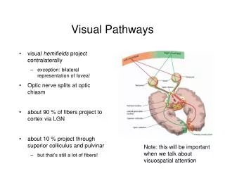
Visual System I – Central Visual Pathways. I. Optic nerve and tract - Axons in the optic nerve cross at the optic chiasm so that each optic tract represents the contralateral visual field - There are three major visual pathways:

E N D
I. Optic nerve and tract • - Axons in the optic nerve cross at the optic chiasm so that each optic tract represents the contralateral visual field • - There are three major visual pathways: • 1) optic tract LGN primary visual cortex (higher visual processing).
2) optic tract superior colliculus (saccades) • 3) optic tract pretectum (pupillary light reflex). • I. Lateral Geniculate Nucleus (LGN) • - Visuotopic map: central retina posterior LGN, peripheral retina anterior LGN
- LGN is made of 6 layers: magnocellular 1,2 and parvocellular 3-6 • - Ipsilateral retina maps to layers 2,3,5 (prime!) and contralateral to 1,4,6.
- LGN neurons have receptive fields that resemble on- and off-center ganglion cells • - Intralaminar cells respond to blue light only, may be responsible for color.
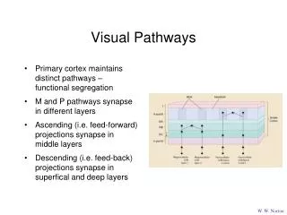
II. LGN to primary visual cortex • - LGN projects dorsally to V1 in the occipital lobe with inferior-superior inversion with respect to visual field • - Columns in LGN project to same part area of V1 (recall that layer 4 receives thalamic input)
- Parvocellular layers 4Cb and 4A in cortex • - Magnocellular layers 4Ca and 4B in cortex • - Intralaminar projects to layer 3
III. Primary visual cortex • - Located in both banks of calcarinesulcus • - Foveal region has high cortical magnification in dorsal V1 • - Only layer 4 is organized in alternating ocular dominance columns
- In V1, input is sent to stellateinterneurons which output via pyramidal cells • - Input and output comes from/goes to LGN, pulvinar, superior colliculus, other cortical areas.
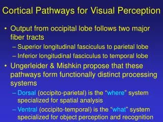
IV. Higher visual areas • - Higher visual areas can be broadly divided into two parts • - Dorsal pathway: spatial position and motion, input primarily magnocellular.
- Ventral pathway: shape, texture, color of objects, input from all layers of LGN • - Secondary visual cortex can be stained to reveal a thick-pale-thin-pale stripe pattern.
- V1 layer 3 thin stripe V2 ventral pathway, V4 • - V1 layer 3 interblobpale stripe V2 ventral pathway, V4 • - V1 layer 4B (magno.) thick stripe V2 dorsal pathway, MT