Download
1 / 51
520 likes | 592 Views
Anatomy of the Female Pelvic Organs. Lulu Al-Nuaim. Aims. To fully understand the anatomy of the female pelvis in terms of bones and tissues, and fetal skull, this would help in explaining the mechanism of Labour To predict and thus prevent postpatum haemorrhae related to the placenta

E N D
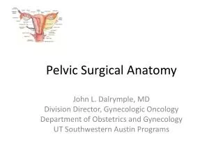
Anatomy of the Female Pelvic Organs Lulu Al-Nuaim
Aims • To fully understand the anatomy of the female pelvis in terms of bones and tissues, and fetal skull, this would help in explaining the mechanism of Labour • To predict and thus prevent postpatum haemorrhae related to the placenta • To understand the major events in fetal circulation; during pregnancy and after birth AL Nuaim
Objectives • Student at the end of session should be able to: • Explain the relationship between pelvic organs • Comprehend the normal organs • Understand the relationship between the female pelvis (Bones& Soft Tissue) and fetal skull, in order to understand the mechanism of labour AL Nuaim
Objectives Student at the end of session should be able to: • Understand the major variant in the fetal circulation than that of the adult • Know the significance of ductusvenoususductusanteriousus and the first breath. • Explain the changes that occur after birth. • Familiarize yourself with the placental structure. • Know the significance of placental and umbilical cord inspection after birth • Differentiate between the different types of placental abnormalities and their significance AL Nuaim
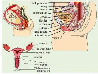
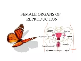
The Vulva external organs of the female Include: - • Mons veneris • Labia majora • Labia minora • The clitoris • The vestibule The vestibule has six openings: • Urethral meatus • Two skene’s ducts • Vaginal orifice • Two Bartholin ducts. AL Nuaim
The Vulva Bartholin glands - lies on each side of the vagina, in the posterior lower third 1/3 of the interiotus. • Secrete mucus – alkaline AL Nuaim
The vagina A Canal/tube extend from the vulva to the uterus • Runs upwards and backwards • Walls lie in close contact antro-posterior. Speculum examination • The posterior vaginal wall is longer than the anterior 11.5 cm (4.5 in) vs 7.5 cm • Cervix enters the vagina at a right angle. • fornices = four Anterior, posterior, lateral AL Nuaim
The Cervix Forms the lower 1/3 of the uterus • Enter the vagina at a right angle • Barrel shape • 2.5 cm (1 in) long • Two parts: • Supravaginal • Intra vaginal • Cervical os • Internal os • External os • Cervical canal between the internal os and the external os • Transformation zone; squamous-columnar junction. AL Nuaim
The Cervix Supports: • Cardinal ligaments • Pubocervical ligaments • Uterosacral ligaments Late in pregnancy – softer and starts to dilate. In labor: • The longitudinal fibres of the uterus contract and retract pulling upward thus reducing the length of the cervix. • The cervix is made up of fibrous and elastic tissue • Full dilatation marks the end of the first stage of labour. AL Nuaim
The Uterus The uterus lies in the true pelvis. Anteverted (A/V)and anteflexed (A/F)in position. The body of the uterus lies above the bladder. • Size: 7.5 cm length • 5 cm wide • 2.5 cm thick • 50 -75 gm weight Gross structure: • The cervix lower 1/3 • The isthmus • The cavity • The corpus • The cornua. • The fundus AL Nuaim
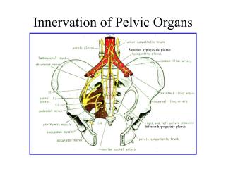
Layers: Endometrium Myometrium Perimetrium - peritoneum Adherent, where??? Loose,??? Blood supply: Arteries: fundus – ovarian artery (aa) Body - uterine artery , directly from internal iliac artery The relationship between the ureter and uterine artery • Uterine artery runs behind the peritonium, cross transverse cervical ligament (Cardinal ligament) then the it passes anterior to and above the ureter 1.5cm from lateral vaginal wall fornix AL Nuaim
Venous: Right ovarian vein - inferior vena cava Left ovarian vein – renal vein Lymph internal and external iliac gland inguinal /Sacral gland AL Nuaim
THE FALLOPIAN TUBEs • Extend from the cornua of the uterus, travels towards the sidewalls of the pelvis. Then turns downwards and backwards. • The tube lies in the upper margin of the broad ligaments • Communicate; superiorly with the uterine cavity, Inferiorly with the perineal cavity • Length 10 cm (4cm) : 3 mm thick • 4 PARTS • Interstitial • Ampulla • Infundibulum • Fimbria AL Nuaim
THE OVARIES • Lie in the posterior wall of the broad ligament at the fibrial end of the fallopian tubes at the level of the pelvic brim. • Size: almond like = 3 x 2 x 1 cm Dull white colour, Corrugated surface • Structure varies with woman’s age. AL Nuaim
The Ovaries • Blood supply – ovarian aa • Ovarian vein • Lymphatic lumbar glands • Nerves ovarian plexus • SUPPORTS They lie in a fossa • Attached to broad ligament – meso ovarian • The meso salpinx is the broad ligament that extend between the fallopian tube and the ovary. • The Fallopion tubes, ovaries and broad ligments are called Adenxa AL Nuaim
Ligaments: • Round ligaments Maintains uterus in A/V + A/F From the cornua of the uterus – pass downwards and insert in the tissue of the labia majora. • Broad ligaments Not true ligament Folds of peritoneum extend laterally from the uterus to the pelvic side walls. • Cardinal ligament transverse perineal muscles • Pubocervical • Uterosacral AL Nuaim
THE NORMAL FEMALE PELVIS The pelvis articulate with the fifth lumbar vertebra above and with the head of each femur in the right and left acetabulum. • The weight of the trunk is transmitted through the pelvis into the legs. • Gives protection to the pelvic organs • The pelvis is the largest bone in the body. Gross structure: Consists of: • 5 fused sacral vertebrae and coccyx • left & right innominate bones • ) AL Nuaim
The Sacrum A triangular shape; The hollow of the sacrum – smooth and concave The alae of the sacrum - give the appearance of wings • The sacral promontary is the centre point of the upper border of the first sacral vertebrae. AL Nuaim
THE COCCYX • 4 Fusesd coccygeal vertebrae • Triangular shape • Articulate with the sacrum • Muscles are attached to its tip. AL Nuaim
Right &Left In-nominate Bones • Each made of 3 separate parts meet in the acetabulum. • Ilium upper part is iliac crest (anterior and posterior, superior iliac crest • Ischiumischialtuberosity , 2 cm above is the ischial spines. • Pubis both meet the pubic body fused by cartilage “symphysis pubis” AL Nuaim
DIVISIONS OF THE PELVIS The brim divides the pelvis into the parts: • The false: lies above the pelvic brim not important in obstetrics • The true: what lies below the pelvic brim. It has a :a brim cavity, and outlet • Forms the curved canal through which the fetus pass during labor. AL Nuaim
The brim or inlet Round in shape • Has eight points as demonstrated • Bounded anteriorly by the pubis • Laterally by illiopectineal lines • Posteriorly by ale and sacral promentary • Widest diameter is, Transverse • True Conjugate ( Anteroposterior diameter) from sacral promentary to upper inner border of Symphysis pubis AL Nuaim
The Pelvic cavity • Extend from the brim above to the pelvic outlet below • The posterior wall 11 cm formed by hollow of the sacrum • The anterior wall is formed by the symphysis pubis and obturator foramen 3.8 cm • The lateral walls sacrosciatic ligamnet and ischial spines • Interspines diameter is shortest diameter AL Nuaim
The pelvic outlet • Obstetrical outlet • The obstetrical outlet The landmarks are: • The lower border of the symphysis pubis • The ischial spines • The sacro-spinous ligament • The lower border of the sacrum. AL Nuaim
Average measurements of pelvis • Brim Antero-posterior = 11.5 cm Transverse = 13.0 cm • Cavity Antero-posterior = 12.0 cm Transverse (I/S) = 10.5 cm Outlet Antero-posterior = 12.5 cm Transverse = 11.0 cm AL Nuaim
Abnormal Pelvis Four Types • Gynecoid Pelvis 50% • Anthropoid 25% • Android Pelvis 20% • Platypelloid (flat 5%
THE PELVIC FLOOR • The outlet of the pelvis is filled with a soft tissue that supports the pelvic and abdominal organs. • It forms as a gutler-shaped structure highest anteriorly than posteriorly. • Three canals with external orifices run through the tissue • The urethra • The vagina • The rectum AL Nuaim
Pelvic Floor • There are six layers of tissue. • An outer covering of skin • Subcutaneous fat • Superficial muscles enclosed in fascia • Deep muscles enclosed in fascia • Pelvic fascia, thickened to form pelvic ligaments • Peritoneum AL Nuaim
Pelvic Floor • Superficial muscles: • Transverse perinei • Bulbo-cavernosus • Ischio-cavernosus • Deep muscles Three pairs of muscles all have their insertion around the coccyx Their anatomical name is levatorani muscles, 5 mm thick • Iliococcygeus • ischiococcygeus • pubo- coccygeus AL Nuaim
perinea body • Lies between the vaginal and rectal canals • Is triangular, the base is the skin and the apex pointing upward each side is 3.8 cm in length • Three layers of tissue 1. outer covering of skin 2. superficial pelvic floor • bubo-cavernous • transverse perinei 3. deep pelvic floor muscle. Episiotomy, types, indications, AL Nuaim
FETAL SKULL • Skull is divided into regions • The vault. • The face. • The base • Bones: Bones are separated by suturs • Two frontal bones • Two parental bones • One occipital bone AL Nuaim
Suture, an area of membrane which has not ossified • Lambdoidal suture • Sagittal suture • Coronal suture • Frontal suture • Anterior fontanelle, diamond in shape where sagittal and frontal sutures meet • Posterior fontanelle, where lambdoidal and sagittal sutures meet. AL Nuaim
AREAS OF THE SKULL • Vertex between anterior and posterior fontanelles • Occipital bone • Mentum: the chin • Frontal bone Malposition Malpresentation AL Nuaim
CIRCUMFERENCES OF THE FETAL SKULL • The engaging Diameter in a well flexed head: suboccipito-bregmatic In Vertex presentation 9,5 cm • The engaging Diameter in a Deflexed head (partly extended) OccipitoFrontal 10,5 cm In Occipito posterior Position AL Nuaim
DIAMETERS OF FETAL SKULL • Bipareital 9,5 cm • Suboccipital-bregmatic 9,5 cm vertex presentation • Occipito frontal 10,5 cm • Mentovertical brow presentation • Submento-bregmatic face presentation AL Nuaim
Effect of Labour and delivery • Moulding • Caput succedaneum • Cephalhaematoma, AL Nuaim
The placenta • Structure of the mature placenta • Maternal surface lies next to the uterus on inspection, chorionic villi are arranged in lobes/cotyledons – 20 in number – 200 lobules. • The groove separating the lobes are sulci • dark – red color, rough surface • Fetal surface, faces the baby. Bluish gray colour, smooth, shiny surface. • Umbilical cord inserted in the fetal surface usually in the centre • Blood vessels seen radiating from the cord • The amniotic membranes covers the fetal surface. AL Nuaim
Structure of the mature placenta • Flat, Roughly circular • 22 cm in Diameter • 2cm thick in the centre • Weight: 1/6 of the baby’s weight AL Nuaim
Abnormalities of placental development. • Placenta succenturiata • Placenta circumvallata • Placenta succenturiata/ Placenta velamentosa andVasa previa AL Nuaim
Umbilical cord: • At full term: 40-50 cm long 1.5 cm in diameter • Twisted in appearance • Two umbilical arteries • One umbilical vein • Wharton jelly • Abnormal insertion of the cord • Battledore insertion marginal insertion • Velamentous insertion in the membrane AL Nuaim
Fetal circulation Cardiovascular system Major variant are explained by: • the presence of umbilical-placental circulation and • absence of significant pulmonary circulation. AL Nuaim
The respiratory function of the placenta requires that oxygenated blood be returned via the umbilical vein and into the fetal circulation. • High venous return from the placenta (oxygenated blood O2 saturation 70-80%) through the umbilical vein. • This maintains the right-left shunt through the foramen ovale • Delivers most oxygenated blood to fetal heart and brain. AL Nuaim
Continue • Placenta -umbilical vein- ductus venosus, • Most of the blood into the inferior vena cava (IVC), this mixes with returning non oxygenated blood from the lower limbs and kidney, liver. However, only partial mixing of the two streams. • Most of the oxygenated blood is directed to the crista dividens at the upper end of the inferior vena cava into the right atrium through the foramen ovale and thus into the left atrium and hence to the left ventricles and ascending aorta to be directed to the brain, heart and upper extremities. AL Nuaim
The remainder of the blood from the superior vena cava mixes with that of IVC and passes directly to the right ventricle. • 10% of it goes through the pulmonary artery to the lung. • Most of this enters the systemic circulation via the ductus arteriosus and into the descending aorta beyond the vessels supplying the head, • It supplies the viscera and lower limbs • It then passes into the umbilical arteries (branches of left and right internal iliac arteries) • High pulomary vascular resistance maintains the right-left shunt through the ductus arteriosus. AL Nuaim
At birth • Blood circulation after birth, The closure of the shunts; • Ductus arteriosus • Foramen ovale • Completes the transition of fetal circulation to newborn circulation AL Nuaim