Download
1 / 19
190 likes | 449 Views
Integration of multiple sources of evidence in clinical classification of VUS. David Goldgar University of Utah School of Medicine. What do we mean by a “VUS”?. Sequence variant in a gene with a clearly established role in a given disease

E N D
Integration of multiple sources of evidence in clinical classification of VUS David Goldgar University of Utah School of Medicine
What do we mean by a“VUS”? • Sequence variant in a gene with a clearly established role in a given disease • Usually rare in the general population (or in the clinically tested population) • If pathogenic would be clinically important to the individual carrying the variant • Typically missense, intronic, or in-frame deletions (but could include others)
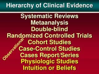
Evidence potentially useful for classification of VUS • Direct: • Co-segregation of VUS with disease in pedigrees • Powerful direct evidence but often difficult to get additional samples from family members. • Co-occurrence (in trans) with deleterious mutations • Only useful if homozygotes/compound heterozygotes are ~embryonically lethal • Distribution of family history of probands carrying a VUS • Indirect: • Severity of amino acid change and evolutionary conservation of wt residue • Effects on protein structure (if known) • Functional evaluation in model systems • Other evidence relevant to cancer susceptibility genes:LOH, pathology, expression array/CGH signatures, MSI
Genetic vs. Functional/Sequence-based Approaches • Genetic approaches normally require multiple observations to be useful; • However, most VUS occur <5 times • Functional and sequence-based analyses can be done (in theory) on any variant • Relationship between functional assay and disease risk typically unknown • If valid relationships could be established, many more VUS could be classified
Select UV Quantifiable Individual or family data Co-occurrence Family History Co-segregation More data LR 1 LR 2 LR 3 Combined evidence (LRi) LR>1000 or LR <0.01 Yes UV classified Validation set for functional and conservation data No Incorporate evidence from conservation and functional data using existing models Initial model Refine model No LR>1000 or LR <0.01 Yes Goldgar et al. AJHG 75:535-44. 2004.
Align-GVGD • An extension of the original Grantham Difference to protein multiple sequence alignments. It uses two variables, GV and GD. • Grantham Variation (GV) • A quantitative measure of the range of variation present at a position in a protein multiple sequence alignment. • GV=0: position is invariant • GV> ~60: non-conservative substitution is tolerated • Grantham Deviation (GD) • A quantitative measure of the fit between a missense substitution and the range of variation observed at its position in the protein. • GD=0: substitution is within the observed range of variation • GD> ~60: substitution is non-conservatively beyond the range of variation • Website http://agvd.iarc.fr 50:0:0
Analysis of rare missense substitutions:Distribution of risk in the GV-GD plane C65 C55 C45 C35 Risk estimates C25 >4.00 C15 3.00-4.00 2.50 to 3.00 2.00 to 2.50 1.67 to 2.00 1.33 to 1.67 1.10 to 1.33 C0 0.90 to 1.10 ≤ 0.90 =0.81 =0.66 =0.29 GD =0.00 GV Tavtigian et al., Human Mutation, (almost) in press 50:0:0
5 x GAL4 bs luciferase Transcription Activation Assay in Mammalian cells (293T) controls - - + + 120 100 LOW RISK 80 %WT (luc activity) 60 40 HIGH RISK 20 0 -Gal4 WT F1695L Y1853X A1830T L1844R P1771L F1662S R1726G R1751Q H1746N M1783T S1613G R1751P M1783L P1859R M1775R Raw data normalized by Renilla luciferase driven by a constitutive promoter. Results from triplicate experiments in which a Gal4 DBD: BRCA1 1396-1863 is co-transfected with the reporter (shown above graph) are plotted as percent of wild type activity. Marcelo Carvalho & Alvaro Monteiro
Estimation of sensitivity and specificity of functional assays (simple approach) • For each variant with functional data, use prior probability based on sequence analysis and log-odds from genetic data to get posterior probability of being pathogenic • Sample each variant as being pathogenic or neutral from posterior distribution • Calculate sensitivity and specificity etc., from this simulated data set • Average over many replicates to get estimated sensitivity/specificity and confidence interval • For Transcriptional Activation assay, estimates were 0.85 (0.67 - 1.0) for sensitivity and 0.65 (0.58 - 0.75) for specificity
The Lyon Meeting on VUS4-5 February, 2008 • Organised by Sean Tavtigian at IARC • Goal to have a highly focused knowledge transfer exercise representing diverse opinions • Representatives from MMR, p16, and BRCA worlds • Assembled expertise: clinical cancer genetics, functional assays, sequence analysis, genetic epidemiogy, etc. • International: US, UK, NL, Australia, France
Series of papers to be written for Human Mutation • Introduction to the series • Genetic variant classification using clinical and epidemiological data • In vitro and ex vivo assessment of functional effects of genetic variants • Splice site alteration assessment • Tumour characteristics as an analytic tool • Integration - the nuts and bolts of combining across data types • Locus specific databases • Clinical utility and risk communication
Issues in Integration • Transferability of results from one kind of mutation to others (e.g., truncating to missense) • LOH, Pathology, Co-occurrence • Choice of appropriate prior probability • Independence of evidence from different sources • Incorporating discrete types of evidence into a probabilistic framework • Combining everything - • Mixture Models via MCMC • Cluster analysis type methods
How to disseminate VUS information to the research and clinical communities • Should research information be separate/different from clinical use? • Qualitative vs. Quantitative information • What is the appropriate place to store this information? • Locus specific databases, e.g. BIC? • clinical databases? • Human Variome database? • All of the above? • How much detail of the evidence should be presented?
Issues in Transfer of Knowledge to Clinical Practice • What are appropriate thresholds for causality and neutrality respectively? • What should be reported? • Only those variants that have been definitively (by above threshold) classified? • Should the ‘current’ odds of causality? • Intermediate discrete categories, e.g., likely deleterious’, probably neutral’? • What if variants confer intermediate risk? Can the methods be adapted to estimate risks? Would it be useful?
Unified Framework for Genetic Testing (including VUS) • Prior probability of an affected proband being a carrier of a pathogenic mutation in gene X based on proband phenotype (including e.g., pathology, MSI, IHC, etc.) and family history and locus heterogeneity; • Could be model based • Add result of genetic testing of proband • wildtype or sequence variant (excluding common polymorphisms) • Add variant specific information • Sequence analysis (A-GVGD, SIFT) • Functional/structural assay if available and quantifiable • Co-segregation analysis if additional family members available to be tested
Unified Framework: Translation into disease risk • From previous information can calculate the posterior probability that the individual carries a pathogenic mutation or wildtype (or a variety of intermediate risks if reliably estimated) • Then disease risk for an at-risk relative of a proband discovered to have variant V is: If V+ : P(V=path)P(D|path)+P(V=wt)P(D|wt; fam hx) If V- : P(V=path)P(Dpop)+P(V=wt)P(D|wt; famhx) • Could be integrated into a single Web-based tool (including sequence, family history, co-segregation, family hx, environmental factors, etc.)
Acknowledgements BRCA2 functional assays: F. Couch, D. Farrugia, M. Argawal, L. Wadum Data Preparation: A. Deffenbaugh, D. Bateman, C. Frye – Myriad Genetics Sequence Analysis: S. Tavtigian, A. Thomas, G. Byrnes BRCA1 functional assays: A. Monteiro, M. Carvalho – Moffitt Cancer Center Statistical Aspects: D. Easton, D. Thompson – Cambridge E. Iversen – Duke University The BIC steering committee; Grants P50CA116201 & R01CA116167 ACS:RSG-040220-01-CCE (FC) and CA92309 (AM)