Download

1 / 12
120 likes | 257 Views
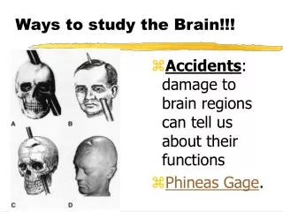
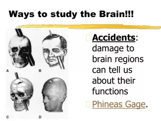
How Psychologists Study the Brain. Accidents. Brain damage due to: Whiplash: aka – diffuse axonal injury Stretching, twisting, damage to the axon Isotropic Stress: high speed change scenarios Car crash, explosions, sky-diving, etc Creates a “pressure wave” or pulse

E N D
Accidents • Brain damage due to: • Whiplash: aka – diffuse axonal injury • Stretching, twisting, damage to the axon • Isotropic Stress: high speed change scenarios • Car crash, explosions, sky-diving, etc • Creates a “pressure wave” or pulse • Causes damage to neurons • Psychologists/Doctors examine: • Loss of Vision • Loss of Hearing • Loss of Memory • Confusion
LesionsDef: cutting the brain, removing portions of the brain • Commonly done in the past • Today: lobotomy • Last resort, experimental surgery • McDreamy • Observe and document changes • Animal experimentation commonly performed • Are correlations reliable and valid? • Harvesting brains of the dead • Concussion victims, dementia, Alzheimer's, schizophrenia, etc
Electroencephalogram (EEG) • Why would you get one? • Sleep Disorder • Having Seizures (epilepsy) • Determine “brain death” • How does it work? • Electrodes attached to the scalp (exterior) • Perform mental function or physical action • Brain waves produced & recorded
Electrical Brain Stimulation (EBS) • How: • Electrodes attached, inserted in the brain (interior) • Charge emitted • Measure results performed by patient • Findings • Specific regions of the brain linked to sensory or motor cortex regions • Ventral Nerves: neurons sending messages to the brain • Epilepsy patients • OCD subjects
Computerized Axial Tomography (CAT) Scanaka – CT scan • Why would you get one? • Car accident • Suspected concussion • Locate blood clot, infection, or tumor • How is it done? • X-ray of the brain • Brain absorbs radiation and computer produces an image • Can be done on any organ
CAT Scans Healthy Brain Scans Abnormal Brain Scans
Magnetic Resonance Imaging (MRI) • Why would you get one? • CAT Scan inconclusive • Looking for a tumor or aneurism • Examine bleeding in the brain • Damage to optic or auditory nerves • How does it work? • Brain produces “energy” • Computer measures radio waves • Looking for abnormalities, “hotspots”
MRI Images A Brain on “MS” Healthy Brain
Positron Emission Tomography (PET) Scan • Why would you have one? • To confirm CAT Scan • To measure blood flow • Diagnose: Alzheimer’s, Parkinson’s, MS, ALS, or Cancer (lymphoma) • How does it work? • Chemical injected (glucose) • Chemical “lights-up” • Image captured PET & CT Machines often are the same
PET Scan Images Schizophrenia Experiment Alzheimer’s Patient PET scans of five normal individuals (left); each row is one person, and each image is a slice from five different levels of the person's brain. The red areas show regions of the brain that are activated when a person performs a memory task. In PET scans of five individuals with schizophrenia (right), each row represents a different person, with comparable slices. Clearly, the patients with schizophrenia do not generate the dramatic brain activity in the circuits of the brain critical to the memory task.