Download

1 / 21
210 likes | 247 Views
Learn about ECG, EKG, vectors, waves, leads, and Holter monitor for thorough heart function assessment. Explore limb and frontal leads, wave patterns, and electrode placement. Essential for medical professionals and students.

E N D
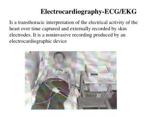
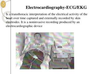
Electrocardiography investigation of heart (ECG). Analysis of ECG
Elecrtocardiogram • It is the method of registration of heart bioelectrical potential from the chest of patient
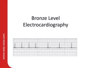
Waves of ECG • 1. P wave – depolarization of atria, precedes atria systole • 2. QRS wave is depolarization of ventricles, precedes ventricular systole • 3. atria repolarization also occurs at QRS • 4. T wave indicates ventricular repolarization
ECG leads • a) Bipolar limb leads. The bipolar limb leads record the voltage between electrodes placed on the wrists and legs. These leads were proposed by Einthoven in 1913. • I lead: left arm (+) - right arm (-); • II lead: left leg (+) - right arm (-); • III lead: left arm (+) - left leg (-). • For recording limb leads we put red electrode on right arm, yellow - on left arm, green - on left leg and black - on right leg. Black electrode has zero potential (ground).
ECG leads • The unipolar limb leads were proposed by Goldberger in 1942. They record voltage between single “exploratory electrode” fro one limb and zero joined electrode from two other limbs. So there are three leads AVR, AVL, AVF. In fact zero electrodes records middle voltage of two limbs. Bipolar limb leads and unipolar limb leads record electrical power in frontal projection.
ECG leads • V1 - in crossing right IV right intercostal space and parasternal line; • V2 - in crossing left IV intercostal space and parasternal line; • V3 - between V2 and V4; • V4 - in crossing V left intercostal space and medioclavicular line; • V5 - in crossing V left intercostal space and anterior axilar line; • V6 - in crossing V left intercostal space and middle axilar line.