Download
1 / 22
230 likes | 395 Views
Chapter 17 - Blood. I. hematocrit. = % of blood volume occupied by erythrocytes average = 45% (average plasma volume = 55%). to determine the hematocrit, a small tube of blood is collected: BLOOD then spun in a centrifuge for 5 minutes: PLASMA BLOOD

E N D
Chapter 17 - Blood Chapter 17
I. hematocrit • = % of blood volume occupied by erythrocytes • average = 45% (average plasma volume = 55%) Chapter 17
to determine the hematocrit, a small tube of blood is collected: BLOOD then spun in a centrifuge for 5 minutes: PLASMA BLOOD erythrocytes are heavier than plasma and are packed in the bottom of the tube plasma is separated from formed elements a special scale is used to determine the relative volume occupied by the erythrocytes compared to total blood volume (see previous slide) Chapter 17
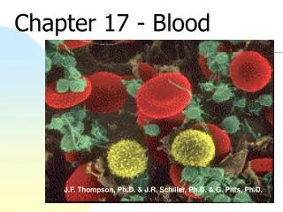
II. erythrocytes • A. physical characteristics • 6 - 8 mm diameter • biconcave discs • more surface area than spherical cells • more flexible and less fragile • no nuclei or organelles • filled with hemoglobin Chapter 17
B. hemoglobin • each molecule of Hb is made of 4 subunits • each subunit contains 1 peptide and 1 heme • normal adult Hb has 2 alpha and 2 beta peptides • each heme contains one • iron atom • the peptide • carries CO2 Chapter 17
C. average lifespan of a RBC = 100 to 120 days • old RBCs are removed from circulation by phagocytes (spleen, liver, bone marrow) • some components are recycled • amino acids • Fe (stored in liver) • the remainder of the heme group is converted to bilirubin and excreted in bile Chapter 17
D. disorders • 1. anemia = reduced ability of blood to carry oxygen • NOT always characterized by low red blood cell count or low hematocrit • a. iron deficiency • b. vitamin B12 deficiency / pernicious anemia caused by lack of intrinsic factor production in stomach Chapter 17
c. aplastic anemia due to destruction of stem cells in bone marrow (radiation, cancer drugs, environmental toxins) • d. hemolytic anemia is caused by destruction of red blood cells • e. hemorrhagic anemia is caused by blood loss • f. genetic disorder causing abnormal hemoglobin Chapter 17
2. polycythemia = high RBC count • a. primary - bone marrow cancer • b. secondary • caused by: • adaptation to increased activity or altitude • dehydration • excess secretion of erythropoietin • effect = increased blood viscosity Chapter 17
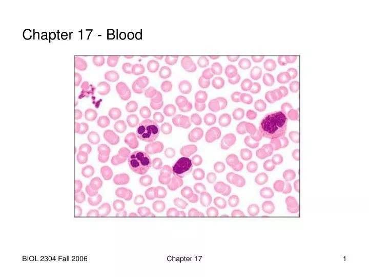
III. leukocytes • A. classification • 1. granulocytes (neutrophils, eosinophils, basophils) • cytoplasmic granules • distorted, inactive nuclei • phagocytic • 2. agranulocytes (lymphocytes, monocytes) • lack obvious granules in cytoplasm Chapter 17
B. characteristics • 1. neutrophils (40 - 70%) • nucleus has 2-6 lobes • cytoplasm stains light purple • phagocytize bacteria Chapter 17
2. eosinophils (1 - 4%) • nucleus has 2 lobes • cytoplasm stains red, orange or dark pink • phagocytize antigen-antibody complexes and fight parasitic worms Chapter 17
3. basophils (0 - 1%) • nucleus has 2 lobes (not visible) • cytoplasm stains dark blue/black • release histamine • and other • chemicals Chapter 17
4. lymphocytes (20 - 45%) • nucleus round, dark, takes up most of cell • cytoplasm seen as thin rim of light blue • about same size as RBCs • functionally divided into 2 categories: • B cells make antibodies • T cells attack foreign, cancer and virus-infected cells Chapter 17
5. monocytes (4 - 8%) • nucleus horseshoe or kidney shaped • cytoplasm pale blue • largest WBC • become macrophages after migrating to c.t. Chapter 17
IV. thrombocytes • fragments of cytoplasm enclosed by plasma membrane • cytoplasm contains secretory granules • involved in hemostasis • form platelet plugs • secrete chemicals that enhance blood coagulation Chapter 17
V. hematopoiesis • occurs in the bone marrow (myeloid tissue) • bone marrow is located inside bones • there are two categories: • red - active • yellow - inactive Chapter 17
A. adult red marrow location: • proximal epiphyses of femur and humerus • axial skeleton • limb girdle bones Chapter 17
B. bone marrow histology • sinusoids (capillaries) • reticular cells, reticular fibers (stroma) • adipose tissue • multipotent blood stem cells (hemocytoblast) • immature blood cells Chapter 17
C. stem cell divides by mitosis to form two daughter cells • one daughter cell becomes the new stem cell • one daughter cell differentiates • this daughter cell becomes the new stem cell • this daughter cell differentiates into a formed element Chapter 17
D. lineages • 1. lymphoid stem cell • lymphocytes • 2. myeloid stem cell • erythrocytes • granulocytes • monocytes • megakaryocytes => platelets Chapter 17