Download
1 / 33
330 likes | 339 Views
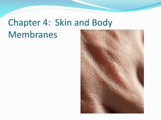
This article discusses the functions and structure of skin and body membranes, including the epidermis, dermis, and various types of epithelial membranes. It also explains the importance of melanin, the rule of nines, and the distinctions between skin cancers.

E N D
DID YOU KNOW?!?: • We have a totally “new” epidermis every 25 to 45 days • Moles and freckles are seen where melanin is concentrated in one spot • Melanin is the natural sunscreen, but excessive sun exposure damages skin causing the elastic fibers to clump leathery skin • Excessive sun exposure weakens the immune system • You can lose up to 7L of body water by sweating on a hot day. • You have more than 2.5 million sweat glands!
Objectives: • List the general functions of each membrane type and give locations • Compare tissue makeup of the major membrane types • Describe functions of the integumentary system • Recognize and label skin diagram as; epidermis, dermis, hair follicle, sebaceous gland, & sweat gland • Describe the layers of the epidermis • Describe the function of melanin • Recognize the importance of the “rule of nines” • Summarize the distinctions between skin cancers • List examples of integumentary aging
Body membranes • Body membranes cover surfaces, line body cavities, form protective (lubricating) sheets around organs • Two categories • Epithelial membranes- Cutaneous, mucous, and serous • Connective tissue membranes- synovial
Epithelial membranes are simple organs • Cutaneous- skin, its superficial epidermis is made up of keratinizing stratified squamous epithelium. A dry membrane that is exposed to air • Mucous (mucosa)- lines all body cavites that open to the exterior such as those hollow organs of the respiratory, digestive, urinary, and reproductive tracts. Comprised of stratified squamousephethelium (mouth) or simple columnar epithelium (rest of digestive tract). These are wet or moist mucosa and are surrounded in secretions
Serous (serosa)- composed of a layer of simple squamous epithelium resting on a thin layer of areolar connective tissue. Lines body cavities that are closed to the exterior (except for the dorsal body cavity and joint cavities) • These membranes occur in pairs: • The parietal layer lines a specific portion of the wall of the ventral body cavity. It folds in on itself to form the visceral layer which cover the outside of the organs in that cavity These layers are separated by a tiny amount of thin, clear fluid , serous fluid, which allows organs to slide easily across the cavity walls w/o friction
Naming serous membranes depends on LOCATION, LOCATON, LOCATION • Peritoneum- serosa lining the abdominal cavity and covering it’s organs • Pleura- lining around the lungs • Pericardium-lining that encloses the heart
Connective Tissue Membranes • Synovial membranes- cushions organs and structures as they move as a result of skeletal/muscular movement • Found at the ends of the fibrous capsules of joints where they provide a smooth surface that secretes a lubricating fluid, synovial fluid, to reduce friction of tendons moving across bone etc…
Integumentary “covering” System (skin, sweat glands, oil glands, hair, and nails) • Functions to protect deeper tissue from: • Mechanical damage detected by cutaneous sensory receptors • Chemical damage prevented by keratinized cells • Bacterial damage prevented by acid secretions of skin • UV damage (melanin produced) • Thermal damage detected by thermal receptors • Drying out prevented by waterproofing keratin and glycolipids Aids in temperature regulation Aids in excretion of urea and uric acid Synthesizes vitamin D from cholesterols (solar powered)
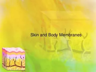
Epidermis- the outer portion is comprised of stratified squamous epithelium that is capable of keratinizing (becoming hard and tough) • Dermis- the underlying layer mostly made up of dense connective tissue • Blister the result of interstitial fluid getting in between and separating the dermis from the epidermis (caused by rubbing)
Subcutaneous tissue (hypodermis)- deep below the dermis it is comprised of adipose tissue. It anchors the skin to the underlying organs. Acts as a shock absorber and insulator, and is responsible for curves of the body.
Strata of the epidermis • From the inside out: • Stratum basale- receives nourishment from dermis via diffusion. Cells constantly dividing million/day • Stratum Spinosum- daughter cells movin on up! • Stratum Granulosum-daughter cells movin on up! • Stratum Lucidum – daughter cells that are full of keratin, flat,clear, and dead! • Stratum corneum- skin region that occurs only where the skin is hairless and extra thick. 20-30 cells thick! Epidermis is avascular (good thing ) Most epidermal cells are keratinocytes (produce protein keratin)
Melanin- a pigment tht ranges in color from yellow to brown to black and is produced by melanocytes that are located primarily in the stratum basale • When sunlight stimulates the melanocytes to produce more of the melanin pigment, tanning occurs. • The stratum basale cells phagocytize (eat) the pigment and it accumulates within them. • Melanin forms a protective pigment umbrella over the superficial side of their nuclei that shields their genetic material from the UV damage
Dermis = “hide” • This dense fibrous connective tissue layer is comprised of two parts: • Papillary layer- the upper layer dermal region, uneven with fingerlike projections (dermal papillae) which indent the epidermis. Contain capillary loops, pain receptors (free nerve endings), touch receptors ( Meissner’s corpuscles), produce ridges of the palms and soles (increase friction) • Reticular layer- deepest skin layer containing blood vessels, sweat and oil glands, deep pressure receptors (Pacinian corpuscles) Found throughout the dermis are: phagocytes to destroy bacteria, collagen fibers for skin toughness and keep skin hydrated, elastic fibers provide the elasticity of young skin.
Age process • As we age the number of collagen and elastic fibers decrease, and the subcutaneous tissue loses fat • The result: skin loses elasticity and begins to sag and wrinkle
Dermis & temp regulation homeostasis • When body temp is high, the capillaries of the dermis become engorged with heated blood and the skin becomes reddened and warm. This allows body heat to radiate from the skin surface • If the environment is cool and body heat must be conserved, blood bypasses the dermis capillaries temporarily, allowing internal body temperature to stay high
Skin color • Three pigments and their amounts contribute to skin color: • Melanin (yellow, reddish brown, or black) • Carotene (orange/yellow) • Oxygen rich hemoglobin (pigment in red blood cells) People who produce a lot of melanin have brown-toned skin. In light-skinned people, the crimson color of the oxygen-rich hemoglobin in the dermal blood supply flushes through the transparent cell layers above
Redness (erythema)- blushing, fever, hypertension, inflammation, allergy • pallor- pale color due to emotional stress, anemia, low blood pressure, reduced blood flow • Jaundice- abnormal yellow skin tone due to liver disorders in which excess bile pigments are absorbed by the blood and deposited • Bruises- reveal sites where blood has clotted in tissue spaces (hematoma) frequent bruising may indicate a vitamin C deficiency or hemophilia
Skin appendages: cutaneous glands, hair, hair follicles, and nails • Cutaneous glands- exocrine glands that release their secretions to the skin surface via ducts. • Two types: • Sebaceous glands • Sweat glands
Sebaceous glands (oil)- found all over skin (except palms and soles) secrete sebum. This is a mixture of oily substances and fragmented cells. Sebum is a lubricant that keeps skin soft, moist, and prevents hair from becoming brittle. Sebum contains bacteria killing chemicals. These glands are very active when male sex hormones are produced in high amounts
Sweat glands-sudoriferous glands are widely distributed is skin ~2.5 million /person • Two types • Eccrine-numerous and everywhere. They produce sweat- a clear secretion that is primarily water, salts, vitamin C, metabolic wastes, lactic acid. Has a slightly acidic pH to prevent bacterial growth. They are supplied with nerve endings to detect high temperature. Sweating through pores is a cooling process due to evaporation. • Apocrine- found in the axillary and genital areas. Larger than eccrine glands and ducts empty into hair follicles. Secretions contain fatty acids and proteins and may be milky or yellowish in appearance. The secretion is odorless however as bacteria on the skin use its proteins and fats, it takes on a musky odor. • These glands begin to function at puberty under the influence of androgens (male sex hormones)
Hair and Hair Follicles • Functions: • Minor protection • Shields eyes • Prevents foreign particle from entering respiratory tract • Minimal insulation Hair follicles produce hair. It is formed by the division of the stratum basaleepethial cells in the matrix (growth zone) of the hair bulb at the inferior end of the follicle. As daughter cells are pushed further from the growth zone, they become keratinized and die. Therefore hair is dead cells and is mostly protein.
Small bands of smooth muscle-arrectorpili- connect each side of the hair follicle to the dermal tissue. • When these muscles contract (cold, fear) the hair is pulled upright, dimpling the skin surface with “goose bumps” • This action allows insulation by trapping air in the fur • Hair pigment is made by melanocytes (yellow, rust, brown, and black) • Hair shaft shape determines hair type and texture
Nail • A scale-like modification of the epidermis that corresponds to the hoof or claw of other animals • Comprised of non living material as the daughter cells produced by the matrix become heavily keratinized
Go to the dermatologist • skin disorders resul from: • Allergies- contact dermatitis • Bacterial infections-carbuncles, impetigo • Viral infections-cold sores • Fungal infections-athletes’sfoot • Autoimmune-psoriasis
Burns-1st, 2nd, 3rd, degree (rule of 9s)- volume of fluid loss is determined by the body surface area that had been burned. Infection is the cause of death in burn victims. Skin is sterile for 24 hours after burn but then bacteria infest the decaying skin matter and thrive. The immune system is suppressed and dehydration, electrolyte loss and protein loss can contribute to circulatory shock
Causes of premature gray hair/hair loss • Genetics- permanent • Emotional crisis • Anxiety • Protein-deficient diets • Chemotherapy • Radiation • Excessive vitamin A • Fungal disease (round worm)
review • Systems in sync pg 125 to understand homeostatic relationships between the integumentary system and the other body systems • Read summary pg 126-127 • Complete all review questions pg 127 • Going further/bonus complete at the clinic responses