Download
1 / 40
400 likes | 418 Views
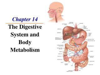
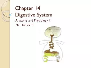
Delve into the intricate structures and functions of the pancreas and liver, including exocrine and endocrine roles, blood circulation, and bile passage. Learn about the diverse cellular components and their contributions in digestion and metabolism.

E N D
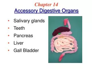
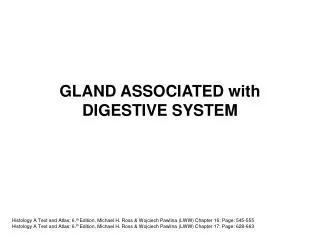
---small gland: fundis gland, small intestinal gland ---large gland: salivary gland, pancreas, liver
---capsule: CT, septa ---parenchyma: /exocrine pancreas /endocrine pancreas
1)exocrine pancreas ---acinus: serous
centroacinar cell: small, pale cell with round or ovoid N, derived from epithelial cell of intercalated duct
---duct: long intercalated duct ---function: secret pancreatic liquid 1-2L/D, PH 7.8-8.4 Digestive enzymes: trypsinogen(胰蛋白酶原) amylase(淀粉酶) lipase(脂酶) chymotrypsinogen (糜蛋白酶原)
2) endocrine pancreas( pancreas islet) ---170.000-200.000, constitute about 1% of total pancreas volume ---75-500 um ---HE: cells arranged into cord with CT rich in fenestrated cap.
a. A cell: • 20%. Large polygonal in shaped, peripheral-distributed • EM: secretory G: large, 190-310 nm, round with dense core • function: secret glucogon(高血糖素) - 29 amino acid residues protein ↓ glycogen→ glucose
b. B cell: • 75%, small, centrally-distributed • EM: secretory G: different diameter, 225-375 nm with one or several dense core • function: secret insulin - 51 amino-acid residues
c. D cell: • 5%, ovoid, fusiform, peripheral-distributed, between A, B cells • EM: -gap junction with A,B cell -secretory G: large, 190-370nm, low-density core • function: secrete somatostatin(生长抑素) to inhibit the secreting of A, B, PP cell
d. PP cell: • EM: secrete granule: small, 110-170 nm • function: secrete pancreatic polypeptide (胰多肽)to inhibit the secreting of pancreatic liquid, movement of viscera and contraction of gall bladder
e. D1 cell: 2-5%, peripheral, irregular EM: small, 140-190 nm, function: secrete VIP (vasoactive intestinal peptide,血管活性肠肽) f. C cell: undifferentiated cell
肝-图 3. Liver
---largest, 2% of body weight ---capsule: DCT, insert into parenchyma to separate the parenchyma into hepatic lobule ---hepatic lobule ---portal area
1) hepatic lobule: basic structural unit ---500,000-1,000,000 ---2 mm long, 1 mm in across D ---polygonal (irregular) ---structure: • central vein • hepatic plate: radiating arranged • hepatic sinusoid
① Central vein: • small vein: endothelium + CT • 45 um • receive the blood from sinusoids
② Hepatic plates ---formed by single layer of hepatocytes
a. hepatocyte: LM: polygonal, 20-30 um eosinophilic N: -large, pale, round, centrally- located -1/4 binucleate
EM: • mitochondria: 1000-2000, 20% total volume • RER: involve in the synthesis of albumin, fibrinogen, clotting factor, lipoprotein and complement protein • SER: contain enzymes- oxidoreductase氧化还原酶 (oxidase, reductase), hydrolase, transterase, synthetase, involve in the formation of bile and the metabolism of adipose, glucose and hormones
Golgi apparatus: involve in -formation of bile -process, condense and storage of proteins -formation of lysosome • Lysosome: involve in phagocytosis activity and metabolism of bilirubin(胆红素) • microbody: -round, 0.2-1.0 um -contain catalase (过氧化氢酶)and peroxidase(过氧化物酶) • inclusions: glycogen, lipid droplet, pigment
b. bile canaliculus: ---cell membrane of adjacent hepatocytes depress to form a tubular system between hepatocytes
---structure: • silver preparation: dark-brown colored network • 0.5-1um • Microvilli • tight junction, desmosome
③ Hepatic sinusoid ---space between hepatic plates ---structure:
9-12 um endothelial cell: fenestrated, gap, plasmalemmal vesicles -liver macrophage (Kupffer cell) -large granular lymphocyte: NK cell
Perisinusoidal space: Disse space - narrow space between endothelial cell and hepatocytes • 0.4 um width; blood plasma • microvilli; RF • fat-storing cell:
fat-storing cell: -irregular, with processes -EM: large lipid droplets, RER, mito, Golgi -function: storage of vitamin A(E,K), synthesis of collagen
The three kinds of different surfaces of hepatocytes ---face adjacent cell each other: 55% ---face the sinusoids: 35% ---form bile canaliculus: 10%
2) portal area ---areas(triangle-shaped or irregular-shaped) where adjacent hepatic lobules meet ---contains CT and several ducts
a. interlobular arteries: • branches of hepatic A • small A: endothelium + 3-4 layers of SM b. interlobular vein: • branches of portal vein • small vein: endothelium + less CT and single SM c. interlobular bile duct: • simple cuboidal or low columnar epi.
3) Blood circulation of liver hepatic A →interlobular A →terminal hepatic arteriole portal V→interlobular V→terminal portal venule → hepatic sinusoid →central vein→sublobular V →hepatic V→inferior vena cava ) {
Blood circulation of liver 门V 小叶间V 终末门微V 血窦 小叶下V 中央V 肝A 小叶间A 终末肝微A 肝V
4) Passage of bile • 肝细胞 胆小管 闰管 小叶间胆管 左右肝管 肝总管 胆囊管 胆囊 或经胆总管到十二指肠