Download

1 / 19
250 likes | 551 Views
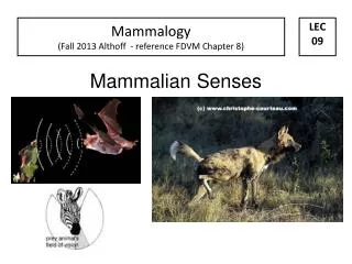
The mammalian eye. By Sze Nga CHAN Cecilia. Content. Structure and function of eye Properties in focusing an image Features of retina Structure of rod cells and cone cells Role of rod cells and cone cells Sensitivity of vision Acuity of vision Effect on rhodopsin.

E N D
The mammalian eye By SzeNga CHAN Cecilia
Content • Structure and function of eye • Properties in focusing an image • Features of retina • Structure of rod cells and cone cells • Role of rod cells and cone cells • Sensitivity of vision • Acuity of vision • Effect on rhodopsin
Features and Functions • Layers • Sclera-tough layer-form the transparent cornea -transparent conjunctiva covers the cornea-transmission-Refraction • Choroid-rich in blood vessels-black in color-prevent internal reflection of light • Retina-photoreceptive-contains rod cells and cone cells
Features and Functions • Aqueous humour-behind the cornea-a clear salt solution • Vitreous humour-behind the lens-jelly-like dark solution • Fovea- contains most of the rod cells and cone cells- light rays are focused onto it • Blind spot-the connection point of optic nerve-no rod cells or cone cells can be found
How to adjust the amount of light goes into the eye? • Strong light-circular muscle contracts-radial muscle relaxes-pupil becomes smaller -less light rays enter the eye Iris is a muscular diaphragm surrounding the pupil
How to adjust the amount of light goes into the eye? • Weak light-circular muscle relaxes-radial muscle contracts-pupil becomes dilate- more light rays enter the eye http://www.youtube.com/watch?v=JadaWSDxBYk&feature=related
How to adjust the focus point? • A near target-ciliary muscles contract-suspensory ligament slack-lens more rounded shape(more convex)-light focused on retina
How to adjust the focus point? • A far target-ciliary muscles relax-pull the suspensory ligament taut-lens flat (pull in)- light focused on retina
Features of retina Three main layers Photoreceptorrod cells and cone cells Bipolar neurones Sensory neurones
Effect on rhodopsin • Light energy is absorbed by retinal • Rhodpsin separate to retinal and opsin • Opsin causes the closure of Na channel • Less inhibitory neurotransmitter is released • Less inhibition of bipolar neurons • Depolarisation • Action potential • Optic nerve
Reformation of rhodopsin • Catalyst • ATP • Trans-retinal + opsin rhodopsin • In iodopsin in cone cells, the same reaction happen • but it breaks less easily and reforms more slowly
Exam question The diagram shows the distribution of rods and cones in the retina of a human eye. Using information in the diagram, explain how: (i) rod cells enable us to see in conditions of low light intensity; Several rod cells to each neuron / bipolar cell/each synapse/convergenceprinciple of additive effect of light striking several rod cells/(spatial)summation; (ii) cone cells enable us to distinguish between objects close together. Each cone connects to a single neurone/no convergence;brain receiving information from each cone cell individually
Reference • http://www.virtualmedicalcentre.com/uploads/VMC/DiseaseImages/2133_eye_anatomy_label_v2_700.jpg • http://upload.wikimedia.org/wikipedia/commons/2/2e/Mona_Lisa_detail_eyes.jpg • http://thebrain.mcgill.ca/flash/d/d_02/d_02_m/d_02_m_vis/d_02_m_vis_1a.jpg • http://www.sapdesignguild.org/editions/edition9/images/accomodation.png • http://www.insightseyecare.net/EyeEducation/retinaNeuronalLayers.jpg • http://cas.bellarmine.edu/tietjen/Laboratories/Eye07.gif • http://4.bp.blogspot.com/_GrFZK0cwrtk/SCXHEojGp5I/AAAAAAAAAB8/7y0hnATxxFg/s400/6a00d41423ae593c7f00d41422a3576a47-320pi.jpg • http://images.ifguk.co.uk/products/609/609-large1.jpg • All information obtained in the internet on 18th April,2010