Download
1 / 38
480 likes | 1.35k Views
Breast Imaging. Olga Hatsiopoulou Consultant Radiologist Royal Hallamshire Hospital Sheffield Breast Screening Unit Sheffield Teaching Hospitals. Screening Breast assessment in symptomatic FT clinics Case studies. Breast Cancer: Why Screen?.

E N D
Breast Imaging Olga Hatsiopoulou Consultant Radiologist Royal Hallamshire Hospital Sheffield Breast Screening Unit Sheffield Teaching Hospitals
Screening • Breast assessment in symptomatic FT clinics • Case studies
Breast Cancer: Why Screen? Improved outcome by treatment during the asymptomatic period Significant impact on public health
Mortality Reduction • 50-69 y.o.: mortality reduction 16-35% • 40-49 y.o.: mortality reduction 15-20% • Lower incidence • Rapidly growing tumors • Dense breasts
Mortality Reduction • Due to detection of cancers at smaller size/earlier stage • Mammographically visible 3-5 years before palpable • Increased detection of DCIS Early stage disease is curable
Diagnostic Accuracy of Screening Mammography • Sensitivity in women > 50 y.o. • 98% fatty breast • 84% dense breasts • Specificity • 82-98%
‘On the positive side, screening confers a reduction in the risk of mortality of breast cancer because of early detection and treatment. On the negative side is the knowledge that she has perhaps a one per cent chance of having a cancer diagnosed and treated that would never have caused problems if she had not been screened.’ Professor Sir Michael Marmot, UCL Epidemiology & Public Health
Triple assessment • Multidisciplinary team approach • Concordance
Concordance of triple assesment P M U B Need for repeat biopsy or clinical core?
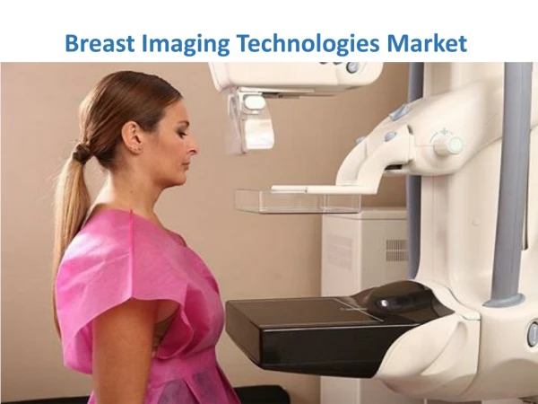
Digital mammography • Quicker to do mammo – almost instant output on monitor • Better penetration of dense breast • Digital manipulation of image
Digital mammography • Proven to be better for younger/denser breasts • Almost eliminates the need for magnification views – can magnify digitally and still have full resolution
Standard view mammography • Cranio-caudal projection (CC) • Medio-lateral oblique projection (MLO)
Calcification • Most are benign and can be dismissed • The goal is to identify new or increasing calcifications or those with suspicious morphology
Malignant microcalcification • Linear, branching casts – comedo • Granular/ irregular – crushed stone • Punctate - powdery
Core biopsy • All solid lumps and M3 MC get a biopsy • Replaces fine needle aspiration in most cases • 14g spring-loaded needle gun • Well tolerated • Main complication is haemorrhage
Core biopsy - histology • Can give grade of cancers and presence of invasion • Can give definitive diagnosis of benign lesions - avoid surgery
Ultrasound vs /stereo biopsy • Ultrasound is used for all lesions visible on ultrasound – quick and accurate • Stereo biopsy is used for lesions not seen on ultrasound –mainly microcalcification (mostly screening women) • Same principle as stereoscopic vision – two slightly different mammographic views allow calculation of depth
Prone biopsy table • Woman lies prone on elevated table with breast dependent through a hope in the table • Biopsy is done from underneath • Access is 360 degrees
VAB • Used with either ultrasound or stereo guidance • Vacuum-assisted biopsy, single needle insertion, larger sample • Allows better non-operative diagnosis, improved calc retrieval, more invasive cancer detection in DCIS
VAB biopsy • 11g, compared with 14g for core biopsy • 8g can be used to remove benign lumps • Slightly greater risk of bleeding • Well tolerated • Can insert clip to mark site in case lesion is totally removed
Why use such a large bore? • A larger sample is more likely to obtain a definitive diagnosis: • DCIS may be upgraded to invasive cancer • ADH may be upgraded to DCIS • Small/difficult lesions are more likely to be adequately sampled • - Therapeutic excision of B3 lesions
Wire localisation • Use U/S or stereo depending on how it is best seen • Aim to get hook through the lesion • Specimen x-ray after excision to confirm lesion remove
LIMITATIONS OF MAMMOGRAPHY • As many as 5 – 15% of breast cancers are not detected mammographically • A negative mammogram should not deter work-up of a clinically suspicious abnormality
FALSE NEGATIVES • Causes • Occult on mammogram (lobular CA) • Finding obscured by dense tissue • Technical • Error of interpretation
RISK OF MAMMOGRAPHY • Average glandular dose from a screening mammogram is extremely low • Comparable risks are: • Traveling 4000 miles by air • Traveling 600 miles by car • 15 minutes of mountain climbing • Smoking 8 cigarettes
Breast MRI • Magnetic resonance imaging is used : • For problem solving • For assessing the extent of lobular or extensive cancers • For screening high risk women - high risk family history and women who have had mantle radiotherapy for Hodgkins’ disease • Pre and post neoadjuvant chemotherapy • For women with implants, to assess integrity
Detecting cancers on MRI • Dynamic scan – bolus injection of Gadolinium and rapid sequence of images • Benign lesions can enhance • Need to create a graph showing pattern of uptake over time • Cancers show rapid uptake and washout
The axilla • Ultrasound • Level one nodes can be very low down • Level three nodes may be best seen from an anterior approach through the pectoralis major muscle
Axillary node levels • Level one: • lateral to lat margin of pectoralis major • Level two: • under pectoralis minor • Level three: • medial and superior to pectoralis minor, up to clavicle
Why scan/ biopsy the axilla? • A pre-operative diagnosis of lymph node metastases will prompt the surgeon to go straight to an axillary node CLEARANCE • A negative axilla on imaging will mean the woman has either: • Sentinel node biopsy • Axillary sampling (four nodes)
Advantages of axillary biopsy • Avoids two operations in women with positive nodes • Alternative is axillary sample at time of WLE, then second operation for clearance
What about PET • Indicated for the complex axilla/ brachial plexus problem • May prove useful for looking for distant mets but not accepted primary method • Resolution and specificity not good enough to look for nodes
Importance of triple assesment • MDT approach • Concordance • Challenges around breast screening • A well informed patient