Download
1 / 49
640 likes | 1.31k Views
Respiratory A&P and Assessment PN 132. ASSESSMENT OF THE RESPIRATORY SYSTEM. Objectives. Identify and define the parts and functions of the upper and lower respiratory system Define common terminology associated with respiratory anatomy, physiology and assessment

E N D
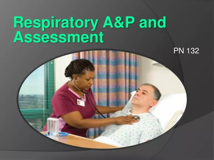
Respiratory A&P and Assessment PN 132 ASSESSMENT OF THE RESPIRATORY SYSTEM
Objectives • Identify and define the parts and functions of the upper and lower respiratory system • Define common terminology associated with respiratory anatomy, physiology and assessment • Identify components of a complete respiratory assessment
Terminology Airway Obstruction Something blocks the airway Prevents air from entering or leaving lungs Anoxia Absence of oxygen Apnea Absence of spontaneous respiration SAS (sleep apnea) Asphyxia Impairment of ventilation and exchange of oxygen and carbon dioxide
Terminology • Bradypnea • Abnormally slow rate of respiration <10 bpm • Cyanosis • Bluish discoloration of the skin caused by a lack of adequate oxygen • Dyspnea • SOB, difficult breathing or labored breathing • Hyperventilation • Abnormally rapid deep breathing • Results in decreased levels of CO2 at cellular level • Hypoxia • Decrease levels of oxygen in inspired gases, arterial blood, or tissues • Just short of anoxia -
Terminology Kussmaul Respirations Deep and labored breathing Respiratory Failure Dangerously low level of oxygen (O2) in the blood OR Dangerously high level of carbon dioxide (CO2) in the blood Tachypnea Abnormally rapid rate of respiration > 20 respirations per minute
The Respiratory System We cannot live without air. Millions of cells in our bodies need a continuous supply of oxygen.
Respiratory System Anatomy and Physiology Respiratory Anatomy Video http://www.youtube.com/watch?v=DCVIEMNPe1E
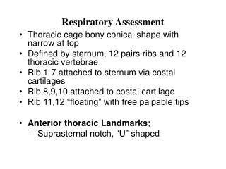
Structures of the Respiratory System Upper Respiratory Tract Nose Pharynx Mouth Larynx Trachea Lower Respiratory Tract Bronchial tree Lungs: alveolar ducts and alveoli
Upper Respiratory Tract Lower Respiratory Tract
The Pleurae Multilayered membranes that are serous and moist Surround and protect each lung Parietal Pleura: outer layer of the pleura Lines the thoracic cavity and forms the sac containing each lung. Visceral Pleura: inner layer of pleura Closely surrounds the lung tissue.
The Pleural Space The space between the folds of the pleural membranes Contains lubricating fluid Prevents friction during respiration. Airtight vacuum Contains negative pressure Keeps the lungs inflated.
The Diaphragm Muscle that separates the thoracic cavity from the abdomen Contracts and Relaxes Phrenic nerve Stimulates diaphragm to contract during respiration.
External Respiration • BREATHING • exchange of oxygen and carbon dioxide • Occurs between the lungs and the environment
Internal Respiration • Exchange of oxygen and carbon dioxide • cellular level • between the alveolus and the alveolar capillaries
CELLULAR RESPIRATION • Exchange of gases within the cells of body organs and tissues. • Oxygen passes from the bloodstream into the tissue cells as carbon dioxide passes from the tissue cells back into the blood stream.
Pulmonary Circulation Superior Vena Cava Pulmonary Arteries Right Lung Left Lung Pulmonary Veins Inferior Vena Cava Aorta
Respiratory System Function • To exchange carbon dioxide (CO2) and oxygen (O2) • To make oxygen (O2) available to the blood stream • So that it can be picked up and used by the cells of organs and tissues in the body • To remove carbon dioxide (CO2) waste from the blood stream
Respiratory Assessment • The respiratory assessment is always included in a patient’s physical exam. • Individuals require more extensive data-gathering - chronic lung conditions - allergic reactions - trauma - recent surgery, etc.
Lung Assessment • SUBJECTIVE • What the patient tells you • OBJECTIVE • What you see and hear
Subjective Assessment • Ask the patient to describe any symptoms he/she is experiencing - shortness of breath - difficulty breathing - cough - orthopnea - pain with inspiration - wheezing, etc.
Subjective Assessment • Data must include details such as - onset - duration - precipitating factors - measures that relieve the symptoms - these may be medications, positioning, oxygen, alternative measures, etc.
Subjective Assessment • Cough • If present, ask for details • Productive/Non-productive • Frequency/sound • If productive, ask for • Color • Amount • Tenacity • Use quotes from the patient whenever possible!
Objective Assessment Observe the patient • Facial expressions when breathing • Chest movement • Quality of respirations - rate, rhythm, depth • Normal Range = 12-20 breaths per minute
Objective Assessment • Observe for • - flaring nostrils • - color of lips and nailbeds • - anxiety on the patient’s face • - skin color and turgor • - equality of breathing on both lungs • - retractions • - Dyspnea • - Orthopnea
Dyspnea and Orthopnea Dyspnea = Difficulty Breathing
Auscultation • Listening for sounds • Auscultate ALL lung fields • Both anteriorly and posteriorly Be sure to warm your stethoscope!!
B = Bronchial BV = Bronchial Vesicular V = Vesicular
Lung Auscultation • The nurse notes the presence of any adventitioussounds (abnormal breath sounds) - wheezes - crackles - pleural friction rub - absence of breath sounds
Normal Breath Sounds Listen: http://www.youtube.com/watch?v=-S8T2JhMrYM
Adventitious Breath Sounds • Abnormal sounds superimposed on breath sounds • Includes: • Crackles (“rales”) • Sibilant Wheezes (“wheezes”) • Sonorous Wheezes (“rhonchi”) • Pleural Friction Rubs
Adventitious Breath Sounds • Crackles: - common on inspiration - interrupted crackling/bubbling sounds - brief, not continuous - can be fine, medium or coarse
Adventitious Breath Sounds • Crackles - Occurs when air is forced through respiratory passages narrowed by fluid, mucous, etc. - Inflammation or infection of the small bronchi, bronchioles, and alveoli - To simulate the sound of Crackles • Take a few strands of hair between your fingers • Hold it up to your ear • Rub back and forth
Adventitious Breath Sounds • Wheezes: Sibilant: - Musical, high-pitched, whistling sounds. - Caused by rapid movement of air through narrowed bronchioles. - May occur during inspiration or expiration - The sound may consist of one or several notes
Adventitious Breath Sounds • Wheezes Sonorous: - Low-pitched, loud, snoring sounds. - Can be heard at any point of inspiration or expiration. - May be continuous
Adventitious Breath Sounds Listen….. http://www.youtube.com/watch?v=_nPi4-ed_Y4#t=19
Adventitious Breath Sounds Pleural Friction Rub • Low-pitched grating or creaking sounds • Heard during both inspiration and expiration • Sound does not originate in the lungs • outside the lung fields • Inflamed pleural surfaces rubbing together during respiration Usually indicates Pleurisy
Adventitious Breath Sounds Pleural Friction Rub This sound occurs when inflamed pleural surfaces rub together during respiration. Listen….. http://www.youtube.com/watch?v=t2QE0O_exAQ
Summary • Defined common terminology associated with respiratory assessment and diagnostic testing • Identified components of a complete respiratory assessment • Identified methods for common respiratory diagnostic testing
Assignment • Read/Review: • PowerPoint Handout • Student Handouts • AHN – Chapter 9 • Pp. 373-379
Next Class • Respiratory Diagnostics and Labs • Understanding ABGs • Look Over • AHN – Chapter 9 • Pp. 379-384