Download
1 / 40
450 likes | 981 Views
Respiratory System Assessment. Chemeketa Community College Paramedic Program. Peggy Andrews, Instructor. Nasal Cavity Oral Cavity Hyoid bone Pharynx Nasopharynx Oropharynx Hypopharynx vallecula. Larynx Thyroid cartilage Cricoid cartilage Arytenoid cartilage Glottic opening

E N D
Respiratory System Assessment Chemeketa Community College Paramedic Program Peggy Andrews, Instructor
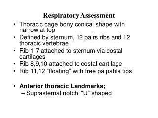
Nasal Cavity Oral Cavity Hyoid bone Pharynx Nasopharynx Oropharynx Hypopharynx vallecula Larynx Thyroid cartilage Cricoid cartilage Arytenoid cartilage Glottic opening Vocal cords Crithothyroid membrane A & P Review- Upper Airway
Trachea Carina Bronchi Left and right mainstem Secondary & tertiary bronchi Bronchioles 22 divisions Respiratory bronchioles Alveoli 1 – 2 cell layers thick Lung parenchyma Pleura Visceral Parietal A & P Review- Lower Airway
Respiratory cycle • Depends on changes in pressure • Inspiration – active process • Expiration – passive process
Measuring oxygen & carbon dioxide levels • Partial pressure of gas • Percentage of mixture’s total pressure • 21% • Diffusion • Movement of gas from higher concentration – lower concent.
Oxygen concentration in blood • Oxygen saturation (SpO2) • PaO2 • 90 – 100 torr normal • Hemoglobin molecule • Carries 4 oxygen molecules • Ventilation/perfusion mismatch • Carbon dioxide concent. In blood
What regulates respirations? • Nervous impulses from the respiratory center • Stretch receptors • Hering-Breuer reflex • Chemoreceptors • Hypoxic Drive
Respiratory rates • Normal - 12 - 20 • Controlled by other factors • Temperature - Emotion • Drugs and medications - Hypoxia • Pain - Acidosis • Sleep • Obstruction • Tongue - most common • Snoring, correct with positioning
Foreign body • May cause partial or complete obstruction • Choking, gagging • Stridor • Dyspnea • Aphonia • Speechless • Dysphonia • Difficulty speaking • Hoarseness
Total Lung Capacity • ~ 6 L • Tidal Volume (Vt) • 500 ml (5 – 7 ml/kg) • Dead space volume • 150 ml in adult male • Minute volume • Vt X RR
Laryngeal spasm and edema • Spasm • Sudden movement/contraction • Most frequently: • Trauma • Aggressive intubation • Post-extubation • Especially if patient semi-conscious
Airway evaluation • Rate • 12-20? • Regularity • Steady pattern • Irregular patterns are significant until proven otherwise
Airway evaluation • Effort • Should be effortless at rest • Changes may be subtle in rate or regularity • Patients compensate by preferential posturing • Upright sniffing • Semi-fowlers • Frequently avoid supine
Some Important Patterns Serious Illness/Terminal DKA Head injury/ICP Resp Center Lesions Paramedic Students
Recognition of airway problems • Respiratory distress • Upper and lower obstruction • Inadequate ventilation • Impairment of respiratory muscles • Impairment of nervous system
Dyspnea may be result of or result in hypoxia • Hypoxia • Inadequate O2 at cells • Hypoxemia • Lack of O2 in arterial blood • Anoxia • No O’s • All therapies will fail if airway inadequate
Visual Clues • S: Pt. c/o sudden onset SOB ~ 2 hrs ago while at rest. PMH: CHF and 2-vessel CABG 1 yr ago. On the usual meds. • O: 67 y/o male Pt CAO PPTE, seated on edge of bed in tripod position. He claims that laying back makes symptoms worse (Orthopnea). Pt. speaks in 2-4 word sentences and frequently needs to be reminded of questions. During assessment, pt becomes increasingly agitated and confused.
What’s your DDX? • What’s your Tx?
Another Sample Pt. What are the clues here? • S: A 62 year old male c/o SOB. Per wife, pt has been unable to sleep and has been having trouble breathing for 4 hours. He has not used his nebulizer treatment because he can no longer hold it to his mouth. PMH: emphysema and asthma.
Our Guy (continued) • O: Pt is CAO Person only, upright in recliner. RR 46, SaO2 64%, Skin pale, cool & moist, with cyanosis around lips, gums, eyes & nailbeds. EKG leads won’t stick to get reading. Lung sounds with minimal air movement in most fields. No wheezes heard. Significant intercostal, supraclavicular, suprasternal and substernal retractions noted on inspiration. Pursed-lip breathing with nasal flaring noted.
DDX? • Tx?
Auscultation techniques • Air movement at mouth and nose • Bilateral lung fields
Palpation techniques • Air movement at mouth and nose • Chest wall • Paradoxical motion • Retractions
Bag-valve-mask • Resistance/changing compliance with BVM ventilations
History • Evolution • Sudden • Gradual over time • Known cause or “trigger” • Duration • Constant • Recurrent • Ease - What makes it better? • Exacerbate – Aggravation of symptoms • Associate - other symptoms (productive cough, etc)
History • Interventions • Evaluations/admissions to hospital • Medications (include compliance and dose) • Ever intubated???
History • Modified form of respiration • Protective reflexes • Cough - forceful, spastic exhalation; aids in clearing bronchi and bronchioles • Sneeze - clears nasopharynx • Gag reflex - spastic pharyngeal and esophageal reflex • Sighing • Increases opening of alveoli • Normally sigh @ 1/min. • Hiccough • Intermittent spastic closure of glottis
Inadequate ventilation • When body can’t compensate for increased oxygen demand or maintain O2/CO2 balance. • Many causes • Infection • Trauma • Brainstem injury • Noxious or hypoxic atmosphere • Renal failure • Multiple symptoms • Altered response • Respiratory rate changes
Supplemental oxygen therapy • Supplemental oxygen therapy • Increases O2 to cells • O2 increases patients ability to compensate • Delivery method continually reassessed
Oxygen source • Compressed gas • Common sizes and volumes • D 400L • E 625L • M 3450L
Calculating Tank Life • Page 386 • Tank Size Factor • 0.16 D Tank • 0.28 E Tank • 1.56 M Tank
Regulators • High pressure • Transfer gas from tank to tank • Cascade System • Therapy regulators • Pressure “stepped down” • Delivery via adjustable low pressure
Delivery Devices • Nasal cannula • Optimal delivery; 40% at 6 Lpm • Indications • Low to moderate enrichment • Long term therapy • Contraindications • Poor respiratory effort • Severe hypoxia • Apnea • Mouth breathing
Delivery Devices • Nasal cannula • Advantages • Well tolerated • Easy to communicate • Disadvantages • Doesn’t deliver high volume/high concentration • % Not guaranteed
Delivery Devices • Simple face mask • Indications • Moderate to high oxygen concentration • 40-60% at 10 Lpm • Advantages • Higher oxygen concentrations • Disadvantages • Beyond 10 LPM does not enhance oxygen content.
Delivery Devices • Partial rebreather • Indications • Contraindications • Apnea • Poor respiratory effort • Advantages • Higher concentrations • Disadvantages • Beyond 10 LPM does not enhance content.
Delivery Devices • Non-rebreather mask • Mask side ports • One-way disc • Reservoir bag attached • 80-95% at 15 Lpm • Indications • Highest O2 content (Non PPV) • Contraindications • Apnea • Poor effort
Delivery Devices • Venturi mask • Mask with interchangeable adapters • Side ports for room air • Highly specific content. O2 • Oxygen humidifiers • Sterile water reservoir for humidifying oxygen • Long term admin. • Desirable for Croup/Epiglottitis/Bronchiolitis • Tracheostomy • Stoma