Download
1 / 47
710 likes | 2.22k Views
Herpesviridae . Properties Enveloped virion, 150 nm in diameter with icosahedral capsid Linear Double stranded DNA genome Replication – nucleus Envelope is acquired by budding through the inner layer of the nuclear membrane Virion produces eosinophilic intranuclear inclusion bodies

E N D
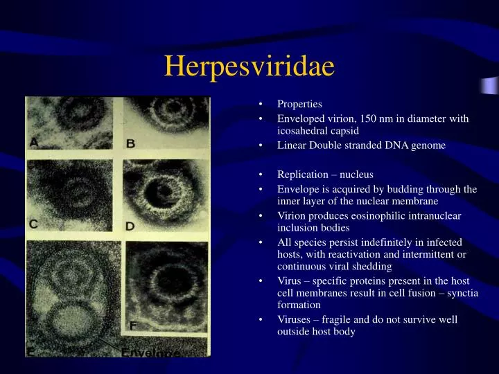
Herpesviridae • Properties • Enveloped virion, 150 nm in diameter with icosahedral capsid • Linear Double stranded DNA genome • Replication – nucleus • Envelope is acquired by budding through the inner layer of the nuclear membrane • Virion produces eosinophilic intranuclear inclusion bodies • All species persist indefinitely in infected hosts, with reactivation and intermittent or continuous viral shedding • Virus – specific proteins present in the host cell membranes result in cell fusion – synctia formation • Viruses – fragile and do not survive well outside host body
Subfamilies and Genera • Subfamily Alphaherpesvirinae • Genus Simplexvirus Human herpesvirus 1 • Genus Varicellovirus Human herpesvirus 3 • Marek’s disease like viruses Gallid herpesvirus 2 • ILTV-like viruses Gallid herpesvirus 1 • Subfamily Betaherpesvirinae • Genus Cytomegalovirus Human herpesvirus 5 • Genus Muromegalovirus Mouse cytomegalovirus 1 • Genus Roseolovirus Human herpesvirus 6 • Subfamily Gammaherpesvirinae • Genus Lymphocryptovirus Human herpesvirus 4 • Genus Rnadinovirus Saimiriine herpesvirus 2
Pathogenesis and Immunity • Major diseases of most domestic animals except sheep • Many are host specific, but some affect a number of species • Herpesviruses are fragile, not surviving well outside the host • Transmission requires close contact, especially mucosal contact – coitus, licking and nuzzling • Short distance droplet spread is a major means of transmission • Varies from acute inflammatory conditions to persistent infection with periodic or continuous shedding. Some are oncogenic • Virus is shed in oral, nasal, genital secretions • Reactivation is frequently observed in immunosuppressed hosts • Humoral and cell mediated response generated • Neutralizing antibody is directed at enveloped glycoprotein peplomeres – max titer at 14 days post infection • Viral antigens on the surface of infected cells are targets for cell mediated immune lysis
Diagnosis • Demonstration of viral antigens • FAT of tissues or smears • Electron microscopic examination of species • Detection of viral n.a. using PCR • Detection of viral antibodies using ELISA, virus neutralization etc. • Four fold increase in antibody titer – seroconversion • Virus isolation – Herpesviruses can be grown in embryonated eggs or in cell cultures derived form their natural host • Gross and histopathology • Intranuclear inclusions • synctia
Subfamily Alphaherpesvirinae • Grow rapidly, lyse infected cells and establish latent infections in sensory ganglia, primarily • Latent genomes survive in episomal extrachromosomal form but rarely may be integrated into the host DNA • Optimum replication – temps lower than normal adult body temp • Most alpha-Herpesviruses produced localized lesions in the skin, CAN or the mucosae of the respiratory and genital tracts • In neonates, less than 3 months of age are generalized infections with foci of necrosis in multiple organs and tissues –low level maternal antibodies • Mononuclear cell- associated viremia may result in the transfer of a virus across placenta in a pregnant animal – abortion results • Some alpha-Herpesviruses have a wide host range
Infectious Bovine Rhinotracheitis • Bovine herpesvirus 1 – rhinotracheitis, genital infections, conjunctivitis, abortion, generalized dz of neonatal calves, encephalomyelitis • World wide • BHV-1 – one serotype, two subtypes have been described • BHV-1.1 – respiratory • BHV-1.2 – genital • Transmission • Rhinotracheitis, conjunctivitis • Aerosol derived from ocular and nasal secretions • Genital infections – primarily coitius, artificial insemination (semen)
Infectious Bovine Rhinotracheitis • Pathogenesis • Viral spread from initial locus may occur through a mononuclear cell associated viremia • Virus can persist for years after an infection • Latent infectious are sources for new outbreaks • All serotype positive animals are considered potential carriers • Latent virus is reactivated with injections of corticosteriods of other immunosuppressive agents that cause active shedding of the virus • Sites of latency – trigeminal nerve – respiratory form, sciatic nerve – genital form • Clinical features • Respiratory disease – necrotic rhinitis, red nose, dust pneumonia • Feedlot, seen 7-14 days after cattle are introduced to the feedlot • Epidemic outbreaks have occurred in dairy cattle
Infectious Bovine RhinotracheitisClinical Features • High morbidity – 100%, but low mortality <10% • Fever, depression, anorexia, salivation and profuse mucopurulent, nasal discharge • Conjunctivitis may or may not be presents • Diffuse hemorrhages and erosions in the muzzle “red nose”, turbinates, nasal mucosa and gums, necrosis of the larynx • Acute uncomplicated cases last 5-10 days, but most recover within 10-14 day • Death usually occurs as a result of a secondary bacterial bronchopneumonia • Conjunctivitis – occur as primary clinical feature or may accompany rhinotracheitis • Inflamed conjunctiva, edematous and accompanied by profuse mucopurulent discharges • May be unilateral or bilateral • Abortion – may occur with RT disease but can occur up to 100 days after infection • Can occur regardless of the severity of the disease in the dam • Generalized disease of neonatal calves – in utero infection – is often fatal • Genital disease – NO ABORTIONS seen
Infectious Bovine RhinotracheitisClinical Features - vaccine • Cows – infectious pustular vulvovaginitis – 1-3 days after coitus • Pain, dysuria, frequent urination, tail swishing, edematous swelling of the vulva, slight vulvar discharge • Vaginal mucosa is bright red with pustules that may become confluent to form a fibrous pseudomembrane- AT LEFT • Uncomplicated lesions heal between 10-14 days • Bulls – infectious balanoposthitis • Inflammation of preputial lining and glans penis with pustule formation • Control • Modified live • Inactivated virus vaccines • Intranasal safe for all cows, abortion may occur with IM modified live vaccine
Bovine Mammilitis and Pseudo-Lumpy Skin Disease • Bovine herpesvirus 2 – BHV 2 • Mammilitis – worldwide • Pseudolumpy skin disease – endemic in Southern Africa • BHV-2 only causes Mammilitis in most countries • Transmission – arthropods and trauma to the skin – Carrier status in recovered animals • Clinical features – Pseudo-lumpy skin disease (see left) • Mild fever followed by skin nodules suddenly appearing over entire body • Healing without scar formation, is complete within a few weeks
Bovine Mammilitis Bovine herpesvirus 2 – NO VACCINE • Clinical features – Mammilitis • Ulcerative lesions – teats and udder • Healing occurs in 3-4 weeks • Reduced milk yield – 10% occurs due to difficulty to milking the affected cows and intercurrent mastitis Control – for Mammilitis and pseudolumpy skin disease • Isolation of infected animals, control arthropods
PseudorabiesAujeszky’s disease – Mad itch • Worldwide disease • Primarily of swine, some domestic animals • Porcine herpesvirus 1 – one serotype • Variations in virulence • May survive in the environment for a few hours to 2-3 days • Rabies is the most sensitive lab animal to pseudorabies infection • Hosts • Adult pigs are the primary hosts and reservoirs - Principal source of virus for a variety of secondary hosts • Secondary hosts – ruminants, dogs, cats, horses, feral animals etc.
PseudorabiesPorcine herpesvirus 1 • Transmission • Spread of the virus from swine to other species and swine is the most important factor in the transmission of pseudorabies • Swine – virus shed in nasal secretions, saliva and milk • Some may shed continuously in their nasal secretions • Licking, biting, aerosols, ingestion of infected carcasses and contaminated water and feed serve as means of transmission • Dogs and cats • Ingestion of infected pig carcasses and rodents • Ruminants • Direct contact with pigs, oral and nasal routes
Pseudorabies in swine • Pathogenesis – following oral or nasal infection, virus replicates in the oropharynx • Virus travels to the various cranial nerve ganglia, medulla and pons via the axoplasm within 24 hours • No viremia • Virus spread continues within the CNS, ganglioneuritis at many sites, nonsuppurative meningoencephalitis and perivascular cuffing • Clinical Features • Weaned, Growing and Adult Swine • IP = 30 hours • Sneezing, coughing fever, profuse salivation, constipation and vomiting • Pigs are listless, depressed, incoordinated, circling and convulsions followed by mortality in less than 2% • Pruritis – dominant feature for secondary hosts – RARE IN PIGS • Piglets born to nonimmune sows: • Sneezing, coughing, encephalitis, prostration and death. Death may occur within 24 hours following parturition. Mortality rate approaches 100% • Maternal antibody is protective therefore, disease in piglets born to recovered or vaccinated sows is less severe with recovery as the outcome
Pseudorabies in Swine – Porcine herpesvirus 1 • Nonimmune pregnant sows – SMEDI • Up to 50% may abort • Infection less than 30th day of gestation results in death and resorption of embryos • Infection late in pregnancy – mummified, macerated, stillborn, weak and normal swine • Less than 20% of sows aborting are infertile on the next breeding BUT do eventually conceive
Pseudorabies in Swine • Clinical features • Weaned, growing and adult swine – IP – 30 hours • Sneezing, coughing, fever, profuse salivation, constipation and vomiting • Pigs listless, depressed and uncoordinated, circling, convulsions, followed by death <2% • Piglets born to nonimmune sows • Sneezing, coughing, encephalitis, prostration and death • Death may occur within 24 hours of parturition • Mortality rate approaches 100% • Maternal antibody is protective, disease in piglets born to vaccinated sows or immune sows is less severe
Pseudorabies in Secondary Hosts • In secondary hosts the disease is characterized by a severe local pruritis with a high mortality • Cattle – MAD ITCH • Intense pruritis – Cattle may become frenzied. Pruritis may not be observed if the virus is acquired via inhalation • Progressive involvement of the CNS • Paralysis of the pharynx, salivation, forced respiration • Death from respiratory failure, occurs within a few hours or may take as long as six days • Dogs and cats • Dogs – frenzy associated with pruritis, mandibular paralysis and pharynx with hypersalivation. Unlike Rabies – dogs are NOT aggressive • Cats – disease is so rapid that pruritis may not be observed
Pseudorabies in Secondary Hosts Porcine Herpesvirus 1 - vaccine • Diagnosis – • History – pig contact in recent past with clinical signs – death following intense pruritis • Virus isolation: • Pigs – CNS, tonsils, lungs and liver • Secondary hosts – CNS, lungs of cattle • FAT staining of frozen tissue sections – brain and tonsils • Serology – Detection of antibodies using serum neutralization, ELISA, latex agglutination tests • Control – • *******************Pseudorabies is a reportable disease ***************** • Vaccination of swine in endemic areas – do not prevent infection or the establishment of latent infection by the wild type virus • Both the modified live virus and inactivated vaccines are used • Gene-deleted vaccine – A pseudorabies vaccine in which the thymidine kinase gene and a glycoprotein gene have been removed is currently available
Equine herpes virus 1 Equine Abortion virus – EHV 1 • EHV – most virulent equine herpesvirus and is associated with “abortion storm”,respiratory disease, perinatal foal disease and encephalitis in horses. • It is the most important cause of viral abortion in horses. It is antigenically related to equine herpesvirus 4. EHV – is endemic and equine populations worldwide • Transmission – inhalation of infectious aerosols, direct or indirect contact with nasal discharges, aborted fetuses, placenta or placental fluids • Pathogenesis – following inhalation of the virus replicates in the epithelial cells of the respiratory tract and rapidly localizes in the regional lymph nodes within 2- 3 days of infection
Equine Herpesvirus 1 – Equine abortion virus • EHV –1 has predilection for circulating leukocytes and endothelial cells • Pregnant mare – • Mac-associated viremia results in infection of the endothelial cells of blood vessels within the uterine wall, other organs and the developing fetus resulting in abortion • Most abortions occur during the LAST TRIMESTER, but can occur as early as the fourth month of gestation. • Prior to abortion, the mare may show signs of respiratory tract infection. • Reproductive efficiency is not compromised • Encephalitis – EHV-1 • Associated with resp. disease and abortion. • Characterized by vasculitis of microvasculature in the CNS, followed by hypoxic degeneration – ischemic – and hemorrhage in adjacent neural tissue • Vasculitis may result from virus – antibody immune complexes – immune complex hypersensitivity – since virus isolation is almost impossible
EHV-1Equine Abortion virus - vaccine • Encephalitis continued… • Severity can vary from slight hind limb in coordination of transient nature • Quadriplegia and recumbency resulting in death • Some recover completely, others suffer permanent damage • Perinatal foal mortality – • Fatal generalized disease accompanied by respiratory distress due to interstitial pneumonia • Rhinopneumonitis – EHV-1 – occasionally causes problems • Control – • An inactivated vaccine is available for EHV-1 to prevent abortion. It is administered during the 5th , 7th and 9th months of pregnancy. • Immunity is short lived – 2-4 months
Equine Herpesvirus 4 - vaccine • EHV-4 – respiratory disease but has been recovered from individual cases of abortion • Distribution – worldwide • Transmission – inhalation of droplets from infected horses and older carrier horses that shed, following reactivation of latent infection • Clinical features – • Mainly in foals over 2 months, weanlings, yearlings • Fever, anorexia, profuse serous nasal discharge later becoming mucopurulent • Most foals completely recover • If there is intercurrent infection, stress, overcrowding, young foals may suffer a fatal bronchopneumonia • Partial immunity, older animals, infections are sub clinical • Control – combined inactivated EHV-1 and EHV-4vaccines are used to control respiratory disease and abortion. • Short lived immunity
Hemorrhagic Disease of PuppiesFading puppy syndrome • First found in 1965 – USA • Highly fatal, generalized hemorrhagic disease of puppies under four weeks of age • Worldwide • Etiologic agent – Canine herpesvirus 1, one serotype • Transmission – newborn puppies can get in utero from passage through the birth canal, from contact with infected littermates, from oronasal secretions of the dam, or rarely fomites • Older dogs – contact with saliva, nasal secretions and urine of infected dogs or puppies, VENEREAL TRANSMISSION
Hemorrhagic disease of puppies • Pathogenesis – puppies • Initial replication occurs in tonsils and nasal epithelium, followed by mac-associated viremia and virus replication in endothelial cells - VIREMIA • Large ecchymotic hemorrhages and necrosis are observed in multiple organ systems - all • Neonatal puppies – infected less than one week of age most unsusceptible to fatal generalized infections • Puppies older than three weeks of age at the time of infection are relatively resistant and generally develop mild or unapparent infection • CHV-1 – optimal temp 33 degrees – 2-4 weeks when thermoregulatory center develops – hypothalamus • Will not see after 4 weeks of age!!! • The more severe the hypothermia, the more rapid the course of the disease • Bitch – viruses multiply in utero and via birth canal • Horizontal and vertical transmission via nasal discharges • 24-48 hours they die
Fading puppy disease- no vaccine • Clinical features • Puppies – 3-8 days, course 1-2 days • Painful crying, abdominal pain, anorexia, dyspnea, soft, odorless yellowish green stool. • NO FEVER • Meningoencephalitis occurs in oronasally infected puppies • Virus may travel up the nerve axons ot the CNS – under normal circumstances, puppies generally die from systemic illness before neurologic signs are manifest. • Older dogs • Mild respiratory infection – rhinitis and pharyngitis • Vaginal or preputial discharge, focal nodular lesion of the vagina, penile and preputial epithelium. Persistence of virus in latent form • Control • Low incidence of severe disease in pups less than 20%, and mild nature of disease in older dogs has resulted in no vaccine; isolation and keeping infected pups separate for 2-3 weeks • Reduced hypothermia – by providing heating whelping box or putting under infrared lamp • Isolation of affected bitch and her litter. Surviving pups will be infective for 2-3 weeks
Feline Rhinotracheitis • Feline herpesvirus 1 – 1957 • One of the 2 most common causes of acute respiratory dz. • In kittens – frothy salivation, ulcers in cornea common, prolonged fever, hypersalivation – dehydration, NO VIREMIA – abortion due to dehydration • Associated with stress, immunosuppression • Distribution – worldwide • Prevalence of antibody in colony cats is over 70% • Household cats – less than 50% • Etiologic agent – feline herpes virus 1 – one serotype • Herpes corneal ulcer • Calicivirus – lingual ulcers • Chlamydia – conjunctivitis – see gyimah chart • Hosts – all members of Felidae • Transmission – FRV shed in ocular, nasal and oral secretions
Feline Rhinotracheitis • Natural routes of infection for FRV are nasal, oral and conjunctival • Recovered cats carry the virus for some time, intermittent shedding and can transmit the disease to susceptible cats • Pathogenesis – replication in the mucosae of the nasal septum, turbinates, nasopharynx and tonsils • Clinical features – IP – 2-6 days, but may be longer if exposure rate is low • Neonates – • Sneezing, coughing, profuse serous nasal and ocular discharge may be mucopurulent, profuse frothy salivation, dyspnea, anorexia, weight loss and fever • Keratitis – punctuate corneal ulcers, oral rare • Kittens less than 4 weeks, extensive rhinotracheitis and associated bronchopneumonia from secondary bacterial infection – MAY BE FATAL
Feline Rhinotracheitis • Older kittens – older than six months, mild or sub clinical disease • Pregnant queen – abortion around the 6th week of pregnancy may occur, virus has not been found in aborted fetuses • Thought to be due to the debilitating affects of the respiratory disease – dehydration etc. that have a direct effect on the fetus • Pathology – necrosis of epithelia of nasal cavity, pharynx, epiglottis, tonsils, larynx, trachea. Osteolytic changes in turbinate bones may be observed. • Diagnosis – history and clinical signs • Gross histopath – inclusion bodies, synctia • Virus isolation – ocular or pharyngeal swab • Serology – four fold increase in virus neutralizing antibody titer
Feline Rhinotracheitis - vaccine • Differential Diagnosis • Feline rhinotracheitis – hypersalivation, sneezing, keratitis • Feline calicivirus – ulcers of the tongue, palate and pharynx; lameness • Feline pneumonitis – chlamydia psittaci- conjunctivitis, ocular discharges • Control • Inactivated or attenuated live-virus vaccines; alone or in combination with other antigens are available • Genetically engineered vaccine compromised of a gene deletion mutant of feline herpesvirus 1 which gene encoding the capsid protein of feline calicivirus has been inserted is being developed
Dyspnea in pullets Infectious Larygotracheitis – Gallidherpesvirus 1 • ILT is acute, highly contagious disease of chicken • Severe dyspnea, coughing and rales • USA – 1926 • Worldwide • Hosts – chickens and pheasants • Etiologic agent – gallid herpesvirus 1, one serotype although strains of virus differ significantly in virulence • Transmission – introduced into a flock by carrier birds • Mostly via inhalation – occasionally via ingestion • Pathogenesis – severe laryngotracheitis characterized by necrosis, hemorrhage, ulceration and the formation of diphtheritic membranes • Diphtheritic membrane – may form a tube the length of trachea – occluding air flow • Death occurs from asphyxiation
Infectious Laryngotracheitis ILTVaccine • Clinical features – 2-8 days • Most common – chickens 4-18 months • Marked respiratory disease – extension and slinging of the neck during inspiration • Head pressing, resting on breast during exhalation, coughing, rattling • Birds may cough up blood mucus that stains walls and posts • Morbidity – 100%, mortality 20-70% - depending on virulence of the strain • Diagnosis – • FAT staining of smears and tissues • Virus isolation – nasal mucosa • Chorioallantoic membrane of embryonated eggs – stunted embryos which die 2-12 days post inoculation • GHV-1 grows well in various cultures • Serology – detection of neutralizing antibody using pock or plaque reduction assays
ILT – Gallid herpesvirus 1 • Control- • Vaccination of replacement pullets, 6 weeks of age or older, in endemic areas using modified live-virus vaccine • Complete depopulation and disinfection of premises have been used to eradicate the disease
Mareks diseaseGallid herpesvirus 2 – serotype 1 • Split paralysis – one leg in front and one behind • One of the most important diseases worldwide • Synonyms – range paralysis – neurolymphomatosis • Hosts – primarily chickens, pheasants, quail and game fowl and also susceptible • Etiologic agent- gallid herpesvirus 2, two serotypes have been identified • Serotype one – virulent strain – and serotype 2 avirulent strain – strains of serotype 1 vary in virulence and in the types of of lesions they produce • Virus slowly cytopathic and remains highly cell associated – mac associated – so that cell free infectious virus is almost impossible to obtain, except in dander from feather follicles • Cutaneous and ocular lymphomatosis at left
Marek’s disease • GHV-2 contains V-onc genes that resemble those in avian retroviruses • Oncoproteins coded by v-onc genes produce a polyclonal T lymphocyte tumor – lymphomatosis • Transmission – • Inhalation of chicken dander • Many healthy birds are lifelong carriers and shed virus however it is not transferred in the egg • Pathogenesis – • Subclinical infection with virus shedding is common • Initial replication occurs in the epithelial cells of the respiratory tract, resulting in a mac-associated viremia • Virus dissemination results in productive infection of various lymphoid organs and tissues • By 2nd week of infection, a persistent cell-associated viremia is followed by TRANSFORMATION OF THE T LYMPHOCYTES • Lesions in Marek’s disease result form the infiltration and in situ proliferation of T lymphocytes • Lysis of nonlymphoid cells by the virus results in a marked inflammatory response
Marek’s disease • Clinical features • Progressive disease with overlapping signs • Outbreaks seen in chicks 2-5 months of age • Mortality may reach 80% • Neurolymphomatosis – classic Marek’s • Enlargement of one or more peripheral nerve trunks. The nerves may be THREE TIMES the normal diameter, show LOSS OF STRIATIONS, EDEMATOUS, GRAY OR YELLOW IN COLOR • Enlargement of nerves is usually on ONE SIDE • Lameness, droopy wings, splayed legs – one held forward and one backward, incoordination, and LIMBERNECK are commonly observed • Visceral and nervous system form will always be present; May or may not have skin or eye form ; Paralysis of the neck muscles, wings – limberneck • Splayed appearance
Marek’s diseaseGallid herpesvirus 2 • Visceral lymphomatosis – Lymphocytic tumors are present in the heart, liver, kidney, and spleen etc. • Ocular lymphomatosis - Graying of the iris – Gray eye – see 1st slide of set – of one or both eyes resulting from T cell infiltration. • There is partial or total blindness • Cutaneous lymphomatosis - Plucking the feathers reveals round, nodular lesions in the skin • Productive infection of the epithelial cells at the base of feather follicles is associated with the release of cell-free virus • Diagnosis – • History, age, clinical signs, and gross postmortem findings are adequate diagnosis • Detection of Viral antigen - FAT • Detection of antibodies – AGID, IFA and virus neutralization tests • Virus isolation – Buffy coat or spleen cells • Cell cultures, CAM or yolk sac routes of embryonated hen’s egg
Marek’s diseaseGallid herpesvirus 2 - vaccine • Control – • Vaccination is the main method of control • An avirulent turkey herpesvirus – heterologous vaccine – and the avfirulent serotype type 2 strain have been used as vaccines. • An attenuated serotype 1 vaccine is also available • The perferred vaccine is the avirulent TURKEY HERPESVIRUS VACCINE because the virus infects birds productively, yielding a high antigenic dose
Subfamily betaherpesvirinaeinclusion body rhinitis - piglets • Synctia formation, inclusion bodies, but not temperatures • Individual viruses have a restricted host range • Slow growing, cell lysis does not occur until several days after infections, they are associated with chronic infections lasting several months before clinical recovery • Infected cells – cytomegaly – cytomegalovirus • Mainly in latent form in secretory glands – salivary – kidneys, lymphoreticular tissues, mac and other tissues • Betaherpesviruses are associated with continuous viral excretion • Inclusion body rhinitis - Endemic worldwide • In a given herd, 90% may be infected • Etiologic agent – porcine herpesvirus 2 – one serotype • Highly cell associated
Subfamily betaherpesvirinae inclusion body rhinitis • Transmission – mainly through inhalation • Transplacental transmission in susceptible sows • Pathogenesis – virus replicates in the mucosa of the nasal passages and the URT • Clinical features – disease seen in 2-10 weeks old • Most severe in piglets less than 3 weeks of age • Sneezing, coughing, serous nasal and ocular discharges, depression, • Discharge thickens – mucoid – and blocks nasal passages • Interferes with suckling, resulting in rapid weight loss and death • Transplacental infection is associated with mummification, stillbirths, neonatal death and runt pigs – SMEDI • Older pigs > 4 months, sub clinical infection • Pathology – widespread petechiae and edema thoracic cavity and SQ tissues
Inclusion body rhinitis – NO VACCINE • Basophilic intranuclear inclusion bodies and cytomegaly are seen in numerous cells • Diagnosis – clinical signs, gross and histopathology • Virus isolation form nasal mucosa, lung and kidney • Detection of antibodies using ELISA and IFA tests • Control – • Good management and abstention from introducing new stock during the mating period and first month of pregnancy
Subfamily Gammaherpesvirinae • Different members of the subfamily replicate in T or B lymphocytes- lymphotrophic • Some cause lymphoid tumors e.g. Burkitt’s lymphoma and nasopharyngeal carcinoma in humans • Some members are slowly cytopathic for epithelial cells, causing death without production of virions • Some gammaherpesviruses are shed continuously from epithelial surfaces • Latency occurs in lymphoid tissue
Malignant Catarrhal Fever- corneal opacity • MCF – sporadic but highly fatal disease of domestic and exotic ruminant sp. • Infects the lymphoid tissue and epithelial cells of the respiratory and GI tracts • Sheep- associated in Europe and American • Africa – wildebeest, deer • Etiologic agent – Africa – alcelaphine herpesvirus 1 – bovine herpesvirus 3 • First isolated from wildebeest • Highly cell associated virus • American/European form – ovine herpesvirus 2, serologic evidence of a close antigenic associate between the American strain and the African strain
Malignant Catarrhal Fever- hemorrhages zebra stripes • MCF – reported in feedlot cattle and North America in the absence of contact with sheep – minor epidemics – identity and source of virus in this third form is unknown • Hosts – cattle, deer, rabbits and buffalo • Reservoirs – wildebeest, hartebeest, topi (Africa), sheep (USA), • Unapparent infections occur in the reservoirs • Transmission – • African form – close contact between cattle and wildebeest, during calving • AHV-1 occurs in nasal and ocular secretions of young wildebeest in a cell free state • American form – close contact between cattle and sheep during lambing • Cattle have CELL ASSOCIATED VIRUS, but not cell-free virus, in secretions, and this may explain the noncontagious nature of MCF when contact occurs with MCF- affected cattle – dead end hosts
Malignant Catarrhal Fever • Malignant Catarrhal Fever has been reported in N.A. feedlot cattle without sheep present • The cause of infection is not known • Hosts: cattle, deer, rabbits and buffalo • Reservoirs – wildebeest, hartebeest, topi (Africa), sheep (USA) Unapparent infections occur in the reservoirs • Transmission – African form. Close contact between cattle and wildebeest, especially during calving. AHV-1 occurs in nasal and ocular secretions of young wildebeest in a cell-free state. • American form – Close contact between cattle and sheep during lambing • Cattle have cell-associated virus, but not cell-free virus, in secretions, and this may explain the noncontagious nature of MCF when contact occurs with MCF affected cattle – DEAD END HOSTS
Malignant Catarrhal Fever • Clinical Features – • Morbidity low – usually only 1-2 cows/herd show clinical signs • Mortality – VERY HIGH • Peracute form – • Characteristic signs and lesions may not develop, since the disease lasts only for 1-3 days • Severe inflammation of the oral and nasal mucosa and hemorrhagic gastroenteritis • Acute form – IP – 2-8 weeks, three weeks average • Fever, depression , profuse nasal and ocular discharges, generalized lymphadenopathy, and extensive mucosal erosions of the GIT – bloody diarrhea – and respiratory tract • Bilateral ophthalmia – photophobia, corneal opacity that begins peripherally and progresses toward the center of the eye. May lead to blindness - common
Malignant Catarrhal Fever • Diagnosis • History and clinical signs – especially bilateral ophthalmia • Virus isolation – African form • Serology – Seroconversion using serum-virus neutralization test • Buffy coat cells, spleen cells, thyroid and lymph node • Cell culture – calf thyroid monlayer cells - Syncytia formation, intranuclear inclusion bodies • Immunity – rare bovine that recovers from MCF may be immune for life - ??? • Control – separate cattle from sheep and wildebeest • Incidence is just too low to develop a vaccine • Others Gammaherpesviruses – • Equine herpesvirus 2 – Respiratory disease, conjunctivitis, and corneal edema in foals • Equine herpesvirus 5 and • Bovine herpesvirus 4 – pathogenicity unknown