Download
1 / 17
170 likes | 316 Views
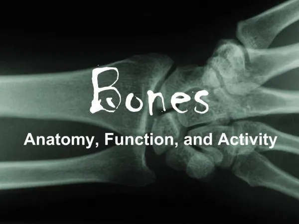
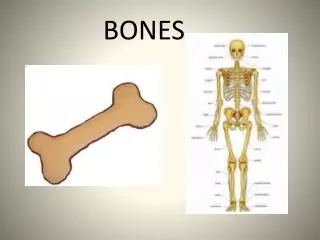
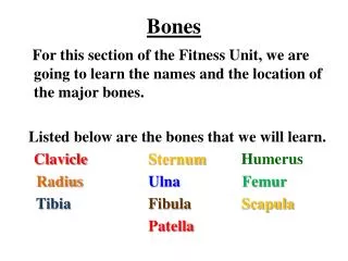
BONES. Functions Provides framework for form and shape of the body (Prevents soft tissue from collapsing ) Provide points of attachment for muscles (Articulation: allowing bone movement relative to one another ) Protection of vital organs

E N D
Functions • Provides framework for form and shape of the body (Prevents soft tissue from collapsing) • Provide points of attachment for muscles (Articulation: allowing bone movement relative to one another) • Protection of vital organs • Storage areas for mineral salts and fats (Ca, P, Na, K) • Blood Cell production
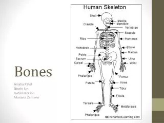
Macroscopic Structure • Diaphysisis a hollow cylinder of compact bone containing yellow bone marrow (fat storage) in the centre • Epiphysescontain spongy (cancellous bone) in centre. Large spaces in spongy bone are often filled with red bone marrow. (Red blood cell production) • Periosteum: dense white fibrous covering • Articular Cartilage: cartilage covering each epiphysis.
Microscopic Structure • Bone is a connective tissue (cells separated by large amounts of non-cellular matrix) with inorganic salt deposits. (increase strength / rigidity) • Composed of bones cells (osteocytes) • Haversian System/ Osteons (compact bone): • Projections from bone cells enter canaliculi and make contact with adjacent cells (Material transfer) • Haversian/Central canal contains at least one blood capillary. (may also contain nerves / lymph capillary) • Haversiansystem runs parallel to long axis of bone (adds strength) • Lamellae: layers of bony matrix • Lacunae: pockets (spaces) between lamellae
Microscopic Structure • 2. Trabeculae(spongy bone): • Irregular arrangement of thin, bony plates. • Contains bone cells but not in concentric layers • Nerves and blood vessels pass through irregular spaces in matrix
Microscopic Structure • A connective tissue • Perichondrium: fibrous protective layer • Only external blood vessels (in perichondrium), relies on diffusion through matrix. • Consists of protein fibres (Collagen) which are embedded in Protein-carb complex (chondrin). • Chondroblasts: immature cartilage cells in the spaces in the matrix (produce matrix which will eventually surround it) • Chondrocytes: mature cartilage cells which are inside a pocket (lacunae) surrounded by chondrin
CLASSIFICATOIN • Three types (based on thickness of fibres in matrix) • Hyaline: Very fine and very dense for strength. E.g. larynx (part), trachea, bronchi, articular cartilage, nose (part). • Elastic: medium thickness with elastic properties. E.g. ear, larynx (part), epiglottis, nose (part) • Fibrocartilage: thick and less dense, allowing for slight compression ideal for areas which withstand high pressure. E.g intervertebral discs, knee & pelvic joint.
Fixed (Fibrous joints) • No movement occurs between the bones involved. • Held in place by fibrous connective tissue • On impact bone fracture rather that joint damage. • E.g. skull, teeth/jaw • 2. Slightly movable (cartilaginous joints) • Allows very limited movement • Held in place by fibrous cartilage • egsymphysis pubis, vertebrae joints, joints between ribs and sternum • 3. Freely movable (synovial joints) • Amount of movement is limited only by ligaments, muscles, tendons and adjoining bones. • Highly mobile but equally weak
Ball-and-socket joints Spherical head of one bone fits into cup-like head of another • Only occur in two places: shoulder (humerus/scapula) & hip (femur/pelvis) • Hinge Joint Allows movement in one plane only. • Convex surface of one bone fits into concave surface of another • E.g. elbow (ulna/Humerus), wrist (radius/carpals) , knee (femur/tibia), ankle (tibia/tarsals), fingers & toes (phalanges)
Pivot Joint • Rounded, pointed or conical end of one bone articulates with a ring (part bone, part ligament) • E.g. 1st vertebrae (head) / 2nd vertebrae & radius / ulna • Gliding Joint Gliding movement in any direction (back/forth, side/side), limited only by ligaments or bony processes. • E.g. carpals, tarsals, sternum/clavicle, scapula/clavicle
Saddle joint Two saddle shaped joints • Allows side/side and back/forth movements • e.g. thumb (carpal/metacarpal) • Condyloid (ellipsoid) joint Slightly convex fits with slightly concave • Allows side/side or back/forth movements • e.g. radius/carpal, metacarpal/phalanges, metatarsals/phalanges
STRUCTURE OFA SYNOVIAL JOINT • Capsule: surrounding and enclosing the joint (2 layers): • 1) fibrous capsule (outer layer), dense, fibrous connective tissue attached to periosteum . Flexibility allows movement but strength prevents dislocation • 2) Synovial membrane (inner layer) , vascular, loose connective tissue • Synovial Fluid (0.5mL): Secreted by synovial membrane, fills synovial cavity. Lubricates, nourishes & contains phagocytic cells.
STRUCTURE OFA SYNOVIAL JOINT • Articular Cartilage: provides smooth surface for movement • Articular Discs: (in knee - Menisci/meniscus) fibrocartilage extending inward from articular Capsule. Divide synovial cavities into two cavities. • Bursae: little sacs of synovial fluid. Prevent friction between a bone and a ligament/tendon/skin • Accessory Ligaments: hold bones together
Factors keeping bones together: • fit of articulating bones • Strength of bone ligaments • Tension by surrounding muscles
Movement of a Joint: • Flexion: (bending) decreases angle between articulating bones • Extension: (Straightening) increases angle between articulating bones • Abduction: movement away from the body • Adduction: movement towards from the body • Rotation: Movement of a bone around its long axis.