Download

1 / 60
600 likes | 620 Views
Learn about Intrauterine Growth Restriction (IUGR) in primigravida at 38 weeks gestation, including etiology, risk factors, maternal causes, placental factors, fetal causes, uterine causes, diagnosis methods, and management guidelines.

E N D
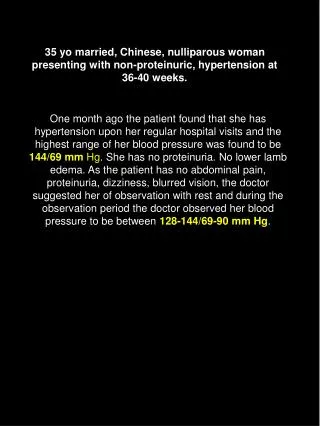
Primigravida, 28 years old with precious pregnancy at 38 weeks gestation. • History of sluggish fetal movments. • O/E: B.P 120/70. • P/A: FH 32 weeks. • Long/ Cephalic. • FHS 95/ min regular.
BPP: 6/10. • Liqour Nil. • Heart rate 103 – 123. • EFW 1.6kg. • P/ V: No liqour or discharge. • Not in labour. • Pelvis adequate.
I.U.G.R INTRA UTERINE GROWTH RESTRICTION
INTRODUCTION • Small sized placenta. • Poor nutrient supply. • Reduced liqour. • Intrauterine fetal death. • Operative and instrumental deliveries.
Perinatal mortality is six to eight times higher. • 40% of the so – called unexplained still birth.
Classification • Very small for gestational age; • Below the 3rd centile. • Small for gestational age; • Below the 10th centile. • Appropriate for gestational age; • 10th to 90th centile. • Above 90th centile ; • Macrosomic
SMALL FOR GESTATIONAL AGE FETUSES. W.H.O Definition ; The term small for gestational age is used to describe a fetus whose growth parameters are below the 10th centile for a given gestational age.
NORMAL SMALL FETUS. • No structural anomalies. • Normal liqour. • Normal umbilical artery doppler study. • Normal growth velocity. • No underlying pathology.
I.U.G.R • Underlying pathology. • Abnormal Umbilical artery Doppler studies. • Further Classification. • Symmetrical I.U.G.R. • Asymmetric I.U.G.R.
SYMMETRICAL I.U.G.R. • Early pregnancy. • Congenital abnormalities. • Infections. • Poor prognosis.
MATERNAL CAUSES • Malnutrition: • Symmetrical I.U.G.R.in 1st trimester • Asymmetrical I.U.G.R. in 2nd trimester
Smoking: • Increased risk is in women who smoke in third trimester and consume more than ten cigarettes per day. • Increased levels of fetal carboxyl hemoglobin.
Alcohol: • 12 fold increased risk of I.U.G.R . • more than 15 units (120g) of Alcohol leads to reduction of 66g of birth weight. • RCOG recommendation in pregnancy. • Causes symmetrical I.U.G.R.
Drugs of abuse: • Heroine and methadone. • TherapeuticDrugs: • B-Blockers , Phenytoin, Anticancer drugs and narcotics
MaternalDiseases: • Cardiorespiratorydiseases. • Anti phospholipid antibody syndrome. • Diabetes. • Chronic hypertension. • Anemia, sickle cell disease, collagen vascular diseases and maternal malabsorption syndrome.
PLACENTAL CAUSES • NORMAL CHANGES IN PREGNANCY • Increase in blood flow from 150ml/min to 600ml/min at term • first layer of trophoblastic invasion in 1st trimester. • 2nd layer of trophoblastic invasion before 20 weeks. • Smooth muscles destruction of spiral arteries.
I.U.G.R. • Second wave of trophoblastic invasion does not occur. • Reduced end diastolic flow velocity. • Decreased oxygen supply to the fetus.
OTHER PLACENTAL FACTORS • Small placental size. • Antepartum hemorrhage. • Thrombosis. • Infarction. • Chrioamnionitis.
Placental cysts. • Chorioangioma. • Placentitis. • Edema.
FETAL CAUSES • Chromosomal abnormalities. • Gastrochisis. • Major cardiac defects. • Fetal infections.
UTERINE CAUSES • Congenital Uterine anomalies. • Large Uterine fibroids.
HISTORY • Age. • LMP. • APH. • Hyperemesis. • Medical history. • Medication. • Obstetric. • Family history.
G.P.E. • B.P. • Pallor. • Dependant edema. • Weight. • Height. • Relevant systemic examination.
Abdominal Examination. • Fundal height measurement. • Sensitivity is 60% - 80%. • Positive predictive value is 20% - 80 %.
Pelvic Examination. • Per speculum examination in cases of ruptured membranes.
Investigations Ultra sound: • BPD, FL and AC . • AC has higher sensitivity and greater negative predictive value. • Type of I.U.G.R. • Serial growth scans. • Four weekly measurement.
Fetal Weight. • Normally 500g at 24 weeks, 1 Kg at 28 weeks, 3.5 kg at 40 weeks. • Formulae • Campbell. • Shephard. • Hadlock.
Liqour Volume • Maximum vertical pool of 2 - 8 cm. • AFI. • After 30 weeks it is between 8 – 25 cm. • AFI of less than 6 cm should be considered seriously.
Doppler • Arterial flow is pulsatile and venous is constant. • Resistance to flow is reflected in the diastolic component. • Reduced EDF indicates high resistance.
Uterine Artery Doppler. • Studied at 20 weeks. • Sensitivity is upto 85%. • Wave form with high resistance or with a notch indicates that the spiral arteries are muscular.
Umbilical Artery Doppler • Performed on high risk mothers. • Diastolic flow is reduced in I.U.G.R. • Absent end diastolic flow is a sign of fetal hypoxia. • Reversed end diastolic flow occurs in severe cases.
Pulsatility Index • Systolic – end diastolic peak velocity • Time average maximum velocity. • Resistance Index • Systolic – end diastolic peak velocity • Systolic peak velocity
Systolic to Diastolic Ratio Systolic peak velocity Diastolic peak velocity
Venous Doppler • Reversed flow • Ductus Venosus. • Umbilical vein pulsations.
Other Tests • Biophysical profile. • CTG. • Karyotyping. • Biochemical markers
Prophylaxis • Low dose aspirin. • Cessation of smoking. • Supplementation. • Bed rest.
Aims of management • Determine the type. • Identify underlying cause. • Deliver at optimum time.