Download
1 / 22
670 likes | 2.43k Views
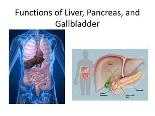
Structure of the liver, gallbladder and pancreas. Sanjaya Adikari Department of Anatomy. Hepatic ducts Common hepatic duct Cystic duct Common bile duct Pancreatic duct Ampulla of Vater Sphincter of Oddi. Ampulla of Vater. Liver. Largest gland in the body Second largest organ

E N D
Structure of the liver, gallbladder and pancreas Sanjaya Adikari Department of Anatomy
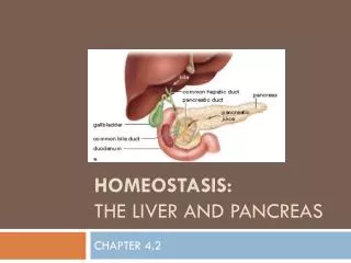
Hepatic ducts Common hepatic duct Cystic duct Common bile duct Pancreatic duct Ampulla of Vater Sphincter of Oddi
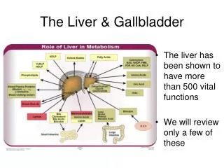
Liver Largest gland in the body Second largest organ Principal metabolic organ 70-80% of its blood comes from portal vein 20-30% of its blood comes from hepatic artery Everything absorbed, except lipids, enter it
Functions of the liver • Fat metabolism • Carbohydrate metabolism • Protein metabolism • Storage • Drug, toxin and alcohol metabolism • Secretion of bile
Liver Pig liver lobules have clear connective tissue boundaries Formed by hexagonal structural units called liver lobules Lobules have a central venule Lobules are surrounded by portal tracts
Liver Human liver lobules do not have clear connective tissue boundaries Lobules are defined by the arrangement of portal tracts around central venule
Structure of a lobule • The main functional cell is hepatocyte • Hepatocytes are arranged in plates • Between hepatocyte plates are sinusoids and bile canaliculi • Sinusoids are lined with fenestrated endothelium • Between the endothelium and the hepatocytes lie the space of Disse
Structure of a lobule • Blood from the branches of hepatic artery and portal vein enter the sinusoids • Blood is drained by central venule, branch of hepatic vein • Bile flow in the bile canaliculi is opposite to the blood flow in the sinusoids • Central venule collects blood from one lobule • Portal tracts supply several lobules around them
Arrangement of portal tracts and central venules Bile ductule
Arrangement of sinusoids and bile canaliculi Bile ductule
Lining cells of sinusoids Three types • Endothelial cells • Kupffer cells • Stellate cells or hepatic lipocytes Stores vitamin A In response to liver injury produces large amounts of collagen causing fibrosis that may lead to hepatic cirrhosis
Kupffer cells These are phagocytic cells Removes aged erythrocytes and particulate matter from circulation
Portal tracts (Portal triads) Branch of the hepatic portal vein Branch of the hepatic artery Bile ductule
Limiting plate The layer of hepatocytes immediately bordering the portal tract is called the limiting plate The limiting plate gets disturbed in diseases like hepatitis
Sinusoids and spaces of Disse Endothelium has no basement membrane Space of Disse lies between the endothelium and hepatocytes Space of Disse drains into the lymphatics of portal tracts
Hepatic acinus Roughly diamond shaped Consists of liver parenchyma centred around a portal tract Acinus is divided into three zones Zone 1 - Receives most oxygenated blood Zone 3 - Receives least oxygenated blood
Gallbladder Bile in the common hepatic duct enters the gallbladder Bile is stored and concentrated in it Capacity is about 100 ml Contracts in response to CCK Bile is an emulsifying agent
Gallbladder Simple columnar epithelium
Pancreas Has exocrine and endocrine components Under the influence of autonomic nerves, gastrin and CCK Secretes enzymes that digest proteins, lipids and carbohydrates Secretes insulin, glucagon, somatostatin, and some other hormones Cells - Insulin Cells - Glucagon Cells - Somatostatin
Pancreatic acini Pyramidal shaped cells Lumen Intercalated ducts