Download
1 / 90
930 likes | 2.51k Views
Lower Back Pain . MS3 Sports Medicine Workshop. Objectives . Review the functional anatomy of lumbo-sacral spine List essential components of a LBP history, including RED FLAGS Describe common causes of LBP Review proper indications for imaging and referral

E N D
Lower Back Pain MS3 Sports Medicine Workshop
Objectives • Review the functional anatomy of lumbo-sacral spine • List essential components of a LBP history, including RED FLAGS • Describe common causes of LBP • Review proper indications for imaging and referral • Review Physical Examination of LS spine • Correlate pathology with pertinent physical findings
“Red Flags” in back pain • Age < 15 or > 50 • Fever, chills, UTI • Significant trauma • Unrelenting night pain; pain at rest • Progressive sensory deficit • Neurologic deficits • Saddle-area anesthesia • Urinary and/or fecal incontinence • Major motor weakness • Unexplained weight loss • Hx or suspicion of Cancer • Hx of Osteoporosis • Hx of IV drug use, steroid use, immunosuppression • Failure to improve after 6 weeks conservative tx
Epidemiology of back pain • Fifth most common reason for all physician visits in US • Second only to common cold as cause of lost work time • 25% of US adults have LBP x1d in last 3 mos • The most common cause of disability in persons under the age of 45
Your patient with LBP has paresthesias in the lateral foot, decreased toe-raise strength, diminished sensation lateral foot, and diminished Achilles reflex. This is suggestive of dysfunction of which nerve root? • L4 • L5 • S1 • S2
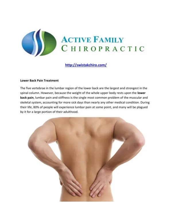
Vertebra • Body, anteriorly • Functions to support weight • Vertebral arch, posteriorly • Formed by two pedicles and two laminae • Functions to protect neural structures
Ligaments • Anterior longitudinal ligament • Posterior longitudinal ligament • Ligamentum flavum • Interspinous ligament • Supraspinous ligament
Ligamentous Anterior longitudinal ligament
Muscles • Spinalis • Longissimus • Iliocostalis • Quadratus lumborum • Ilium to lumbar TPs • Intertransversalis • Interspinals • Multifidus • Erector spinae
Sciatica is defined as… • Pain radiating up the back • Pain radiating to the thigh • Pain radiating below the knee • Pain in the butt
L4 • L5 • S1
PATIENT HISTORY “OPQRSTU” • Onset • Palliative/Provocative factors • Quality • Radiation • Severity/Setting in which it occurs • Timing of pain during day • Understanding - how it affects the patient
Which one is NOT considered a “red flag” of LBP? • History/suspicion of cancer • Age over 50 • Fever or chills • Sciatica
“Red Flags” in back pain • Age < 15 or > 50 • Fever, chills, UTI • Significant trauma • Unrelenting night pain; pain at rest • Progressive sensory deficit • Neurologic deficits • Saddle-area anesthesia • Urinary and/or fecal incontinence • Major motor weakness • Unexplained weight loss • Hx or suspicion of Cancer • Hx of Osteoporosis • Hx of IV drug use, steroid use, immunosuppression • Failure to improve after 6 weeks conservative tx
Onset • Acute - Lift/twist, fall, MVA • Subacute - inactivity, occupational (sitting, driving, flying) • ?Pending litigation • Pain effect on: • work/occupation • sport/activity (during or after) • ADL’s
Other History • Prior h/o back pain • Prior treatments and response • Exercise habits • Occupation/recreational activities • Cough/valsalva exacerbation
Cancer Age > 50 History of Cancer Weight loss Unrelenting night pain Failure to improve Infection IVDU Steroid use Fever Unrelenting night pain Failure to improve Fracture Age >50 Trauma Steroid use Osteoporosis Cauda Equina Syndrome Saddle anesthesia Bowel/bladder dysfunction Loss of sphincter control Major motor weakness Diagnoses & Red Flags
Physical ExaminationMsk Big-6 • Inspection • Palpation • Range of motion • Strength testing • Neurologic examination • Special tests
Approach to LBP • History & physical exam • Classify into 1 of 4: • BAD: LBP from other serious causes • Cancer, infection, caudaequina, fracture • LBP from radiculopathy or spinal stenosis • Non-specific LBP • Non-back LBP • Workup or treatment
What to do aboutPossible BAD Low Back Pain • Cauda Equina: • MRI STAT Neurosurgery consult • Fracture: x-rays • MRI/CT if still suspect • Cancer: x-rays + CRP, ESR, CBC (+/- PSA) • MRI if still suspect • Infection: x-rays; CRP, ESR, CBC, +/- UA
Radiculopathy, Spinal Stenosis • Sciatica (pain below knee) • May have abnl neuro exam • Radiates to leg • Pain worse walking, better sitting (pseudo-claudication)
What to do aboutSuspected Radiculopathy or Spinal Stenosis • Refer to Physical Therapy • Follow in 2-4 weeks for progress • If no improvement by 6-12 weeks • Plain films, MRI, +/- EMG/NCV • Refer for interventions • Epidural steroid injections for radiculopathy
Spondylosis (Osteoarthritis of facet/disk) Spondylolysis/-listhesis Kyphosis/scoliosis Acute lumbar strain Facet pain Discogenic pain Ligamentous pain Causes of “Non-specific LBP”
Management of an acute low back muscle strain should consist of all the following EXCEPT: • X-rays to rule out a fracture • Educate the patient on generally good prognosis • Non-opiate analgesics • Remain active
What to do aboutNon-specific Low Back Pain • Educate patient about expected good prognosis • Advise to remain active as tolerated • Provide analgesics and self-care directions • FU in 2-4 weeks; adjust tx as needed • Don’t do x-rays unless it becomes chronic • WU if no improvement
Renal dz (pyelo, stones, abscess) Pelvic dz (PID, endometriosis, prostate) Gastrointestinal dz (cholecystitis, ulcer, cancer) Retroperitoneal dz Aortic aneurysm Zoster Diabetic radiculopathy Rheumatologic disorders Reiters Ankylosing Spondylitis Inflammatory bowel dz Psoriatic spondylitis Neoplasia (multiple myeloma, metastatic CA, lymphoma, leukemia, spinal cord tumors, vertebral tumors) “Think Outside the Back”
What to do aboutNon-back LBP • WU and tx as appropriate for suspected diagnoses
Diagnostic Studies Radiographs Early if RED FLAGS Symptoms present > 6 weeks despite tx
Diagnostic Studies MRI indications Possible cancer, infection, cauda equina synd >6-12 weeks of pain Pre-surgery or invasive therapy Disadvantages False-positives; may not be causing pain More costly, increased time to scan, problem with claustrophobic patients
Diagnostic Studies Bone Scan indications Adolescent LBP (r/o spondy) SPECT scan Cost ~$300
Diagnostic Studies EMG/NCV r/o peripheral neuropathy localize nerve injury correlate with radiographic changes order after 6-12 weeks of symptoms Pre-surgical or invasive therapy
Lab Studies Indications Chronic LBP Suspected systemic disease CBC, CRP, ESR, +/- UA, SPEP, UPEP Avoid RF, ANA or others unless indicated
Issues specific to CHRONIC LBP(>6 weeks and/or non-responsive) • Evaluation • X-rays, labs • Evaluate for “YELLOW FLAGS” • Management • Medication selection • Interventions
YELLOW FLAGS in Chronic LBP • Affect: anxiety, depression; feeling useless; irritability • Behavior: adverse coping, impaired sleep, treatment passivity, activity withdrawal • Social: h/o abuse, lack of support, older age • Work: believe pain will be worse at work; pending litigation; workers comp problems; poor job satisfaction; unsupportive work env’t
Medications in Chronic LBP • FIRST: Acetaminophen • Second: NSAIDs • If one fails, change classes • Meloxicam naproxen COX2’s • Third: tramadol • Fourth: tri-cyclic antidepressants • Radiculopathy: gabapentin • LOATHE: narcotics
Non-pharmacologic treatments EFFECTIVE NOT EFFECTIVE/ CONFLICTING EVIDENCE BACK SCHOOLS LOW-LEVEL LASER LUMBAR SUPPORTS PROLOTHERAPY SHORT WAVE DIATHERMY TRACTION ULTRASOUND • Acupuncture • Exercise therapy • Behavior therapy • Massage • TENS • Spinal manipulation • Multidisciplinary rehab program
Epidural Steroid Injections • Indicated for radiculopathy not responding to conservative mgmt • Conflicting evidence • Small improvement up to 3 months • Less effective in spinal stenosis
Surgery for Chronic LBP • Most do NOT benefit from surgery • Should have ANATOMIC LESION C/W PAIN DISTRIBUTION • Significant functional disability, unrelenting pain • Several months despite conservative tx • Procedures: spinal fusion, spinal decompression, nerve root decompression, disc arthroplasty, intradiscal electrothermal therapy
Inspection • Observe for areas of erythema • Infection • Long-term use of heating element • Unusual skin markings • Café-au-lait spots • Neurofibromatosis • Hairy patches, lipomata • Tethered cord • Dimples, nevi (spina bifida)