Download
1 / 33
340 likes | 494 Views
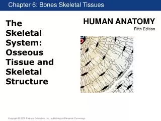
Skeletal Tissue. A. Tissues 1. Cartilage A) 3 types 1) Hyaline cartilage a) Articular cartilage – covers ends of bones. Skeletal Tissue. b) Costal cartilage – connects the ribs to the sternum c) Laryngeal cartilage – forms skeleton of the larynx

E N D
Skeletal Tissue A. Tissues 1. Cartilage A) 3 types 1) Hyaline cartilage a) Articular cartilage – covers ends of bones
Skeletal Tissue b) Costal cartilage – connects the ribs to the sternum c) Laryngeal cartilage – forms skeleton of the larynx d) Tracheal & bronchial cartilage – reinforces passageways of respiratory system e) Nasal cartilage – supports the external nose
Skeletal Tissue 2) Elastic cartilage a) External ear b) Epiglottis 3) Fibrocartilage a) Intervertebral discs b) Menisci of the knee c) Pubic symphysis
Skeletal Tissue B) Cartilage Growth 1) Appositional growth a) New matrix is secreted by chondroblasts onto the edges of the already existing piece 2) Interstitial growth a) Chondroblasts within the lacunae secrete new matrix within the already existing piece
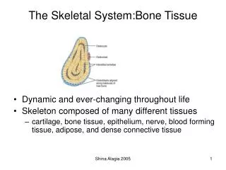
Skeletal Tissue 2. Osseous Tissue (Bone) A) 2 main types 1) Compact – small amount of space between its hard components
Skeletal Tissue 2) Spongy – large spaces between its hard components; known as trabeculae
Skeletal Tissue B) Histology of Bone 1) Compact bone a) Osteon (Haversian system) i) Concentric lamella – circular layers of compact bone
Skeletal Tissue ii) Haversian canal iii) Volkmann’s canals – connect adjacent Haversian canals iv) Lacunae – small gaps at lamellar junctions v) Osteocytes
Skeletal Tissue vi) Canaliculi – connect the lacunae b) Interstitial lamella – between osteons c) Circumferential lamella – around entire outside of bone just under periosteum
Skeletal Tissue 2) Spongy bone a) Not very complex i) composed of several layers of lamella & osteocytes ii) no osteons or blood vessels
Skeletal Tissue C) Bone Development 1) ossification – process of bone tissue formation a) intramembranous ossification – bone develops from a CT membrane i) responsible for most skull bones and the clavicle ii) begins at 8th week of development
Skeletal Tissue b) endochondral ossification – bone develops from a hyaline cartilage model i) all other bones ii) begins by the end of the 3rd month
Skeletal Tissue 2) bone growth in length a) occurs at epiphyseal plates (growth plates) i) chondrocytes in this area are stacked upon one another
Skeletal Tissue ii) cells at epiphyseal end divide rapidly iii) cells at diaphyseal end enlarge, calcify, and ultimately die iv) osteoid replaces calcified cells
Skeletal Tissue b) slows with age until epiphysis and diaphysis fuse i) about 18 for females and 21 for males 3) bone growth in width a) appositional growth i) osteoblasts beneath periosteum (outer surface) lay down new bone tissue ii) osteoclasts beneath the endosteum (inner surface) break down old bone tissue, but usually at a slower rate
Skeletal Tissue 4) hormonal control of bone growth a) human growth hormone (hGH) – from pituitary gland i) controls growth at growth plate b) thyroid hormones (T3 & T4) i) regulate the function of hGH
Skeletal Tissue c) testosterone & estrogen (sex hormones) i) released at puberty ii) cause growth spurt and skeletal changes associated with puberty iii) also induce epiphyseal plate closure; now known as epiphyseal line
Skeletal Tissue D) Bone homeostasis 1) Remodeling a) the ongoing replacement of old/worn bone tissue with new bone tissue b) areas of increased stress can be replaced a couple times a year while some bones never have their tissue completely replaced c) also replaces injured bone
Skeletal Tissue d) cells involved i) osteoclasts – responsible for the breakdown of bone tissue ii) osteoblasts – responsible for production of new bone tissue iii) osteocytes – mature bone cells
Skeletal Tissue f) process – similar to appositional growth i) osteoclasts digest bone tissue by releasing enzymes & acids ii) osteoblasts produce new bone tissue around themselves until surrounded iii) once osteoblast is surrounded it becomes an osteocyte
Skeletal Tissue g) unlike growth in length & width this process occurs in specific areas and is coordinated so as not to alter bone shape 2) Calcium homeostasis a) bone contains almost 99% of the body’s Ca++ stores b) blood levels are very closely regulated i) if they rise too high = cardiac arrest ii) if they drop too low = respiratory arrest
Skeletal Tissue c) nerve action, muscle contraction, enzymes, and clotting all require specific Ca++ amounts to function properly d) bone acts to buffer blood Ca++ levels i) if levels are too high, Ca++ is taken up (new bone tissue is generated) ii) if levels are too low, Ca++ is released (current bone tissue is broken down)
Skeletal Tissue e) regulated by hormones i) parathyroid hormone – from parathyroid gland (a) increases osteoclast activity ii) calcitonin – from thyroid gland (a) increases osteoblast activity
Skeletal Tissue E) Bone repair (of fractures or breaks) 1) Fractures are treated by reduction – realignment of broken bone ends a) Closed reduction – bone ends coaxed back into place by the physician’s hands b) Open reduction – involves surgery; bone ends are secured together with pins or wires
Skeletal Tissue 2) Process of bone repair a) Hematoma formation i) Blood vessels in bone tear and hemorrhage occurs resulting in a mass of clotted blood b) Fibrocartilage callus formation i) Capillaries grow into the hematoma and phagocytic cells invade the area
Skeletal Tissue ii) Fibroblasts, chondroblasts, and osteoblasts migrate to the fracture iii) Fibroblasts secrete collagen fibers and chondroblasts that secrete a cartilage matrix iv) Osteoblasts begin forming spongy bone v) The mass of tissue is referred to as a fibrocartilage callus
Skeletal Tissue c) Bony callus formation i) Osteoblasts and osteoclasts continue to migrate inward and continue to replace connective tissue with bone tissue ii) The tissue is now called the bony callus d) Bone remodeling
Skeletal Tissue 3) Fracture types: a) Simple (closed) – bone breaks cleanly, but does not penetrate the skin b) Compound (open) – broken ends of bone protrude through the tissue and skin c) Comminuted – bone breaks into many pieces d) Compression – bone is crushed (due to porous bone)
Skeletal Tissue e) Depressed – broken bone is pressed inward (skull) f) Colle’s – distal part of the radius breaks g) Transverse – break occurs across the long axis of a bone h) Impacted – broken bone ends are forced into each other i) Spiral – ragged break as a result of excessive twisting of the bone
Skeletal Tissue j) Epiphyseal – break occurring along the epiphyseal plate k) Greenstick – bone breaks incompletely l) Pott’s – malleolus of tibia and/or fibula break
Skeletal Tissue F) Bone Disorders 1) Metastatic calcification – deposition of calcium in tissues that normally don’t store calcium 2) Osteomyelitis – infection of periosteum, medullary cavity and bone 3) Osteoporosis – bone breakdown outpaces bone production
Skeletal Tissue 4) Spina bifida – portions of vertebrae of spinal column fail to form a complete bony arch around the spinal cord 5) Achondroplasia – defective cartilage growth and deficient ability of endochondral bone formation (dwarfism) 6) Acromegaly – delaying of ossification of epiphyseal cartilage (gigantism) 7) Osteotitis – inflammation of bony tissue
Skeletal Tissue 8) Osteomalacia – inadequate mineralization of bone due to insufficient calcium as a result of a vitamin D deficiency (Ricketts) 9) Osteoma – tumor composed of bone tissue 10) Osteotomy – cutting of bone 11) Ostectomy – surgical removal of bone 12) Ostalgia – pain in bone