Download
1 / 32
490 likes | 1.52k Views
HEART DEVELOPMENT. Prof. Saeed Abuel Makarem. Objectives. By the end of the lecture the student should be able to: Describe the formation, site, union, division of the of the heart tube. Describe the formation and fate of the sinus venosus.

E N D
HEART DEVELOPMENT Prof. Saeed Abuel Makarem
Objectives • By the end of the lecture the student should be able to: • Describe the formation, site, union, division of the of the heart tube. • Describe the formation and fate of the sinus venosus. • Describe the formation of the interatrial and the interventricular septae. • Describe the formation of the two atria and the two ventricles. • Describe the partitioning of the truncus arteriosus and formation of the aorta and pulmonary trunk. • List the most common cardiac anomalies.
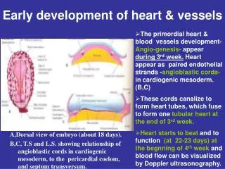
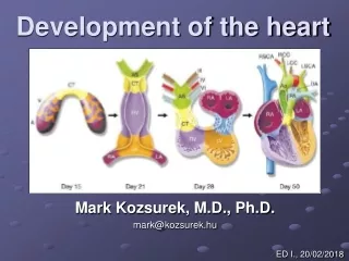
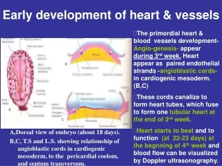
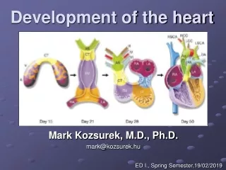
FORMATION OF THE HEART TUBE • The heart is the first functional organ to develop. • It develops from splanchnic mesoderm (cardiogenic area), cranial to the developing mouth and nervous system. • It lies ventral to the developing pericardial sac. • The heart primordium is first evident at 18 days(as an angioplastic cords which soon canalize to form the 2 heart tubes). • After completion of the head fold, the developing heart tubes lie in the ventral aspect of the embryo and dorsal to the developing pericardial sac. • After lateral folding of the embryo • the 2 heart tubes fuse together to form a single endocardial heart tube. • It begins to beat at 22 to 23 days.
Blood flow begins during the beginning of the fourth week and can be visualized by Ultrasound Doppler
Development of the Heart tube • After lateral folding of the embryo, the 2 heart tubes approach each other and fuse in a craniocaudal direction to form a single endocardial heart tube within the pericardial sac.
What is the fate of the Heart Tube? • The heart tube grows faster than the pericardial sac, so it shows 5 alternate dilations separated by constrictions. • These are: • Sinus Venosus. • Truncus Arteriosus. • Bulbus Cordis. • Common Ventricle. • Common Atrium. The endocardial heart tube has 2 ends: 1. Venous end; Sinus Venosus. 2. Arterial end; Truncus arteriosus
U-SHAPED HEART TUBE • Bulbus cordis and ventricle grow faster than the other regions. • So the heart bends upon itself, forming • The U-shaped heart tube, or (bulboventricular loop). bulboventricular loop
Loop formation Or S-Shaped Heart Tube • As the heart tube develops it bends, upon itself: SO, the atrium and sinus venosus become dorsal to the truncus arteriosus, bulbus cordis, and ventricle. • By this stage the sinus venosus has developed 2 lateral expansions, called the 2 horns ( right and left horns) and a body.
Veins Associated With Heart Development Each horn of the sinus venosusreceives 3 veins: 1.Common cardinal 2.Vitelline 3.Umbilical Cardinal vein from the fetal body. Vitelline from the yolk sac. Umbilical from the placenta.
Fate of Sinus Venosus • The right horn forms the smooth posterior wall of the right atrium. • The left horn and the body of the sinus venosus atrophy and form the coronary sinus. • The left common cardinal vein forms the oblique vein of the left atrium.
Right Atrium • The right horn of the sinus venosus forms the smooth posterior part of the right atrium. • Rough Trabeculated anterior part of the right atrium is derived from the primitive or primordial common atrium. • These two parts are demarcated by the crista terminalis internally and sulcus terminalis externally.
Left Atrium • Rough Trabeculated part: derived from the primitive or common primordial atrium. • The smooth part: derived from the absorbed part of the Pulmonary Veins.
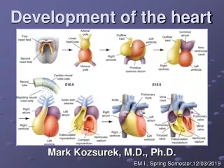
Partitioning of Primordial Heart Partitioning of: 1- Atrioventricular canal. 2- Common atrium. 3- Common ventricle. 4- Bulbus cordis. It begins by the middle of 4th week. It is completed by the end of 5th week.
Partitioning of the atrioventricular canal • Two dorsal and ventral subendocardial cushions are formed on the dorsal and ventral walls of the AV canal. • The AV subendocardial cushions approach each other and fuse together to form the septum intermedium. • Dividing the AV canal into right & left canals. • These canals partially connect the primordial atrium and primordial ventricle.
Partition of the common atrium Septum Primum • A sickle- shaped septum grows from the roof of the common atrium towards the septum intermedium. • So the common atrium is divides into right & left halves.
Ostium Primum • The two ends of the septum primum reach to the growing subendocardial cushions before its central part. • So the septum primum bounds a foramen called ostium primum. • It serves as a shunt, enabling the oxygenated blood to pass from right atrium to left atrium. • The ostium primum become smaller and disappears as the septum primum fuses completely with subendocardial cushions (septum intermedium) to form the interatrial septum.
Septum Secundum • The upper part of septum primum that is attached to the roof of the common atrium shows gradual resorption forming an opening called ostium secondum. • Another septum descends on the right side of the septum primum called septum secundum. • It forms an incomplete partition between the two atria. • Consequently a valvular foramen forms, (foramen ovale).
Fate of foramen Ovale • At birth when the lungs inflated and pulmonary circulation begins the pressure in the left atrium increases and exceeds that of the right atrium. • So the two septae oppose each other. • Its site is represented by the Fossa Ovalis. • The septum primum forms the floor of the fossa ovalis. • The septum secondum forms the margin of the fossa ovalis which is called the limbus ovalis or (anulus) ovalis.
Partitioning of Primordial Ventricle Muscular part of the interventricular septum. • Division of the primordial ventricle is first indicated by a median muscular ridge, the primordial interventricular septum. • It is a thick crescentic fold which has a concave upper free edge. • This septum bounds a temporary connection between the two ventricles called interventricular foramen.
Interventricular Septum The membranous part of the IV septum is derived from: 1- A tissue extension from the right side of the endocardial cushion. 2- Aorticopulmonary septum. 3- Thick muscular part of the IV septum.
Spiral Aorticopulmonary Septum • A spiral septum develops in the truncus arteriosus dividing it into aorta and pulmonary trunk. • So, now the pulmonary artery joins the rightventricle while the aorta joins the left ventricle.
BULBUS CORDIS • The bulbus cordis forms the smooth upper part of the two ventricles. • Right Ventricle: • Conus Arteriosus or (Infundibulum) which leads to the pulmonary trunk. • Left ventricle: • Aortic Vestibule leading to ascending aorta.
Atrial Septal Defects (ASD) • Absence of septum primum and septum secundum, leads to common atrium. • Absence of Septum Secundum
Excessive resorption of septum primum(ASD) Patent foramen ovale
VENTRICULAR SEPTAL DEFECT (VSD) • Roger’s disease • Absence of the membranouspart of interventricular septum. • Usually accompanied by other cardiac defects.
TETRALOGY OF FALLOT Blue Baby • Fallot’s Tetralogy: • 1-VSD. • 2- Pulmonary stenosis. • 3-Overriding of the aorta • 4- Right ventricular hypertrophy.
TETRALOGY OF FALLOT Blue Baby
(TGA) OR TRANSPOSITION OF GREAT ARTERIES • TGA is due to abnormal rotation or malformation of the aorticopulmonary septum, so the right ventricle joins the aorta, while the left ventricle joins the pulmonary artery. • One of the most common cause of cyanotic heart disease in the newborn • Often associated with ASD or VSD. Blue Baby
Persistent Truncus Arteriosus • It is due to failure of the development of the aorticopulmonary (spiral) septum. • It is usually accompanied with VSD.