Download
1 / 12
220 likes | 1.52k Views
DIFFUSION TENSOR IMAGING AND TRACTOGRAPHY. Introduction . Diffusion- random molecular motion also known as Brownian movement Biological systems depend on diffusion for normal functioning like diffusion of metabolites into the cell. . Introduction .

E N D
Introduction • Diffusion- random molecular motion also known as Brownian movement • Biological systems depend on diffusion for normal functioning like diffusion of metabolites into the cell.
Introduction • Diffusion MRI- Produces in vivo images of biological tissues weighted with the local microstructural characteristics of water diffusion. based on • 2 types- DWI and DTI based on isotropy and anisotropy
Isotropy- symmetrical diffusion in all directions • Anisotropy- asymmetrical diffusion in different directions
DWI • Most applicable when the tissue of interest is dominated by isotropic water movement • Eg- grey matter and major brain nuclei • DTI • Most applicable when the tissues have a highly organised structure and hence diffusion is anisotropic because of barriers • Eg- Neural axons- myelin forms important barrier
Terminologies 1) Eigen values- ƛ1, ƛ2, ƛ3- to quantify the diffusion in three orthogonal directions 2) Fractional anisotropy- The anisotropy is expressed relative to the magnitude of diffusion. FA values ranges from 0 to 1( 0- isotropic and corresponds to a perfect sphere 1- anisotropic diffusion and corresponds to ideal linear diffusion) 3) Mean Diffusivity (ADC)- average of the three eigen values
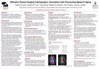
Tractography • A technique used to demonstrate the white matter tracts • Uses DTI sequence. The number of fibres and degree of anisotropy have a direct relationship
Colour coding of tracts • RED- Directions in x axis ( left-right) • GREEN- Y axis ( Posterior to anterior) • BLUE- z axis ( Superior to inferior) Colour- direction of greatest diffusion Brightness- degree of anisotropy
Applications • Traumatic brain injury-DAI • Assessment of the morphology of tracts in lesions adjacent to eloquent areas of brain • Spinal disorders- • Acute and chronic spinal cord compression • Spinal cord tumours- characterisation • AV malformations • Syringomyelia • Alzheimer’s disease
DTI in TBI • Reliable tool for in vivo quantification of white matter microstructural alterations following TBI • They provide valuable information about the axonal integrity and function • Useful for the identifying the early signs of axonal injury in TBI
Studies on DTI state that it is a clinically relevant biomarker in TBI • Tool for revealing the changes in neural tissue during recovery
Also useful in assessment of severity of DAI • Can have a bearing on functional outcome and ? therapeutic impact.