Download
1 / 40
400 likes | 458 Views
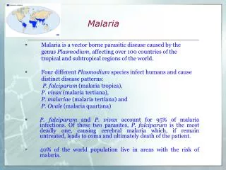
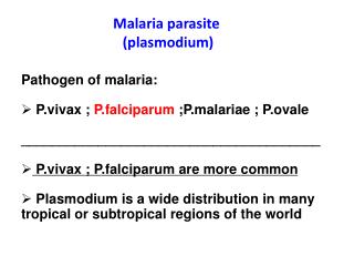
Discover the history, morphology, and clinical features of the Plasmodium parasite causing malaria. Learn about its life cycle, diagnostic stages, and pathogenesis. Uncover the symptoms, incubation period, and complications associated with different species of Plasmodium.

E N D
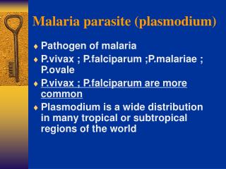
History • Malaria is an old infectious disease. The first documentation about it is at 1500BC. • Until the end of the 19th century, it was commonly thought that malaria was caused by breathing bad air (mal-aria) and was associated with swamps
History • Important application of the knowledge about malaria: W. Gorgas successfully implemented control strategies for malaria and yellow fever during the construction of Panama Canal
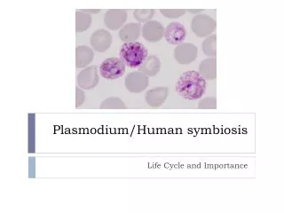
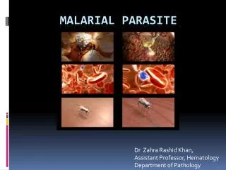
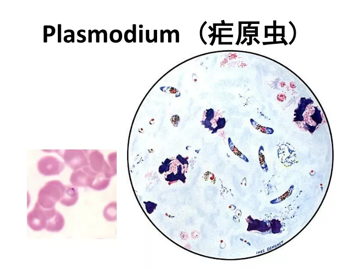
Morphology • Plasmodium is the one-cell parasite, so the basic morphology is a nucleus (chromatin), cytoplasma and cell membrane • Wright or Giemsa stain gives the Cytoplasm – bluish; Chromatin - red to red-purple while the malarial pigments are yellow-brown • There are three stages and six main forms of plasmodium in RBC
Plasmodium in RBC Trophozoites (滋养体期):ring form and developing trophozoites Schizonts (裂殖体期):immature and mature -- merozoites Gametocyte (配子体期): Microgametocytes and macrogametocytes
Trophozoites • Fig. 1: normal red cell; Figs. 2-5: ring stage parasites (young trophozoites)
Ring form trophozoites • Ring like plasma with one nucleus at one side Thin blood film (Giemsa stained)
Mature trophozoites (amoeboid form) • The plasmodium grow with pseudopods, more cytoplasma and malarial pigment presented in the plasma • Red blood cell enlarged and became pale with Schüffner‘s dots(薛氏小点)
Schizonts • Figs.: increasingly mature schizonts
Giemsa staining compact nucleus, usually at edge of the parasite scattered pigment granules The gametocyte is completely filling its host cell Macrogametocyte (female gametocyte) of P.v
Microgametocyte (male gametocyte) of P.v • Giemsa staining • large nucleus at the center of the cell • scattered pigment granules
Macrogametocyte of P. f • The crescent-shaped gametocytes of P. falciparum are very distinctive, but tend to only appear late in the infection • Compact nucleus, red, usually at the center of the cell • Malarial pigments around the nucleus
Microgametocyte of P. f • Sausage-shaped with two blunt end • Large nucleus at the center • Sometimes hard to distinguish from the female gametocytes
exo-erythrocytic stage— merozoites in liver cells
Development in the vector Gametocytes zygote oocyst sporozoites
Life cycle • Infective stage:sporozoites • Transmission • female Anopheles mosquito • transfusion • transplacental • needle stick • Pathogenic stages:erythrocytic stage • Diagnositic stages:erythrocytic stage
Clinical features Incubationperiod(潜伏期) • Time interval between the mosquito bite and the onset of the clinical symptoms • Time for the sporozoite reaching liver and entering • Duration of the development in the liver • Time of development in the RBC to produce sufficient erythrocytic merozoites to cause clinical symptoms • P. falciparum 7 to 27 days • P. vivax 8 to 31 days • P. malarie 18 to 40 days • P. ovale 16 to 18 days
Clinical features Typical malaria paroxysm(发作) • Malarial paroxysm -symptom for erythrocytic phase • RBCs rupture releasing merozoites, malarial metabolites, endotoxin in blood • shaking chill(寒战) followed by fever (高热)(2-20 hrs) • profuse sweating when fever breaks(出汗退热) • repeated characteristic cycle for each species P. falciparum 36-48hrs P. vivax 48hrs P. malariae 72hrs P. ovale 48hrs
Recrudescence versusrelapse • Recrudescence (再燃)is the return of the symptom of malaria after apparent cure, which is due to a sudden increase in what was a persistent, low-level parasite population in the blood. • The periodic increase in numbers of parasites results from a residual population persisting at very low levels in the blood after inadequate or incomplete treatment of the initial infection. • The asymptomatic situation may last for as long as 53 years.
Recrudescence versus relapse • Victims may suffer relapse (复发)after apparent recovery of malaria. This is caused by the long prepatent sporozoites (LPPs) or hypnozoites which remain dormant in the hepatocytes for an indefinite period. • P. vivax and P. ovale will develop hypnozoites in the liver responsible for relapse whilepatients infected withP. falciparum and P. malariae have no phenomenon of relapse • All four types of plasmodium can maintain low level of parasitemia and account for the recrudescence of the disease
Clinical features • liver & spleen damage of long-term • complications from P. falciparum • Anemia • tropical splenomegaly syndrome • cerebral malaria - 50% of deaths • coma and renal failure due to tubular necrosis
Diagnosis • Travel history to endemic area & presence of symptoms, e.g., fever and chills • Thick smears - hard to read • Thin smears - see RBC morphology and parasite characteristics • serology - helps rule out fever of unknown origin • DNA probes- too slow & costly esp. in field
Diagnosis Thick blood smear Thin blood smear
Treatment • Quinidine(奎尼丁), chloroquine(氯喹), primaquine(伯氨喹), pyrimethamine(乙胺嘧啶), artemisinin(青蒿素), sulfadoxine(磺胺), mefloquine(甲氟喹), tetracycline(四环素), proguanil(氯胍) • affect parasite in different ways depending on stage when administered • different species react differently • emergence of drug-resistant malaria
Prevention and control • Personal protection- netting, repellents, etc. • mosquito control or eradication - not easy • avoid sharing needles • develop vaccines - not currently available • select proper treatment for species and morphological forms
summary • Relapse • Recrudescence • Typical malaria paroxysm • How to reduce the morbidity and mortality of malaria?