Download
1 / 151
1.55k likes | 1.84k Views
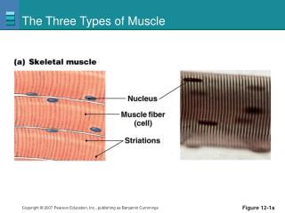
Three Types of Muscle Tissue. Skeletal muscle tissue: Attached to bones and skin Striated Voluntary (i.e., conscious control) Powerful Primary topic of this chapter. Three Types of Muscle Tissue. Cardiac muscle tissue: Only in the heart Striated Involuntary

E N D
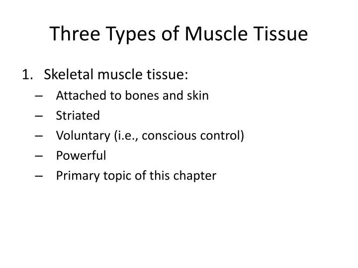
Three Types of Muscle Tissue • Skeletal muscle tissue: • Attached to bones and skin • Striated • Voluntary (i.e., conscious control) • Powerful • Primary topic of this chapter
Three Types of Muscle Tissue • Cardiac muscle tissue: • Only in the heart • Striated • Involuntary • More details in Chapter 18
Three Types of Muscle Tissue • Smooth muscle tissue: • In the walls of hollow organs, e.g., stomach, urinary bladder, and airways • Not striated • Involuntary • More details later in this chapter
Epimysium Epimysium Bone Perimysium Endomysium Tendon Muscle fiber in middle of a fascicle (b) Blood vessel Fascicle (wrapped by perimysium) Endomysium (between individual muscle fibers) Perimysium Fascicle Muscle fiber (a) Figure 9.1
Features of a Sarcomere • Thick filaments: run the entire length of an A band • Thin filaments: run the length of the I band and partway into the A band • Z disc: coin-shaped sheet of proteins that anchors the thin filaments and connects myofibrils to one another • H zone: lighter midregion where filaments do not overlap • M line: line of protein myomesin that holds adjacent thick filaments together
Thin (actin) filament Z disc H zone Z disc Thick (myosin) filament I band A band Sarcomere I band M line (c) Small part of one myofibril enlarged to show the myofilaments responsible for the banding pattern. Each sarcomereextends from one Z disc to the next. Sarcomere Z disc Z disc M line Thin (actin) filament Elastic (titin) filaments Thick (myosin) filament (d) Enlargement of one sarcomere (sectioned lengthwise). Notice the myosin heads on the thick filaments. Figure 9.2c, d
Longitudinal section of filaments within one sarcomere of a myofibril Thick filament Thin filament In the center of the sarcomere, the thick filaments lack myosin heads. Myosin heads are present only in areas of myosin-actin overlap. Thick filament Thin filament Each thick filament consists of many myosin molecules whose heads protrude at opposite ends of the filament. A thin filament consists of two strands of actin subunits twisted into a helix plus two types of regulatory proteins (troponin and tropomyosin). Portion of a thick filament Portion of a thin filament Myosin head Tropomyosin Troponin Actin Actin-binding sites Active sites for myosin attachment Tail Heads Actin subunits ATP- binding site Flexible hinge region Myosin molecule Actin subunits Figure 9.3
Sarcoplasmic Reticulum (SR) • Network of smooth endoplasmic reticulum surrounding each myofibril • Pairs of terminal cisternae form perpendicular cross channels • Functions in the regulation of intracellular Ca2+ levels
T Tubules • Continuous with the sarcolemma • Penetrate the cell’s interior at each A band–I band junction • Associate with the paired terminal cisternae to form triads that encircle each sarcomere
Triad Relationships • T tubules conduct impulses deep into muscle fiber • Integral proteins protrude into the intermembrane space from T tubule and SR cisternae membranes • T tubule proteins: voltage sensors • SR foot proteins: gated channels that regulate Ca2+ release from the SR cisternae
Part of a skeletal muscle fiber (cell) I band A band I band Z disc H zone Z disc Myofibril M line Sarcolemma Triad: T tubule • • Terminal cisternae of the SR (2) Sarcolemma Tubules of the SR Myofibrils Mitochondria Figure 9.5
Sliding Filament Model of Contraction • In the relaxed state, thin and thick filaments overlap only slightly • During contraction, myosin heads bind to actin, detach, and bind again, to propel the thin filaments toward the M line • As H zones shorten and disappear, sarcomeres shorten, muscle cells shorten, and the whole muscle shortens
Z Z H A I I 1 Fully relaxed sarcomere of a muscle fiber Z Z I A I 2 Fully contracted sarcomere of a muscle fiber Figure 9.6
Requirements for Skeletal Muscle Contraction • Activation: neural stimulation at aneuromuscular junction • Excitation-contraction coupling: • Generation and propagation of an action potential along the sarcolemma • Final trigger: a brief rise in intracellular Ca2+ levels
Events at the Neuromuscular Junction • Skeletal muscles are stimulated by somatic motor neurons • Axons of motor neurons travel from the central nervous system via nerves to skeletal muscles • Each axon forms several branches as it enters a muscle • Each axon ending forms a neuromuscular junction with a single muscle fiber
Myelinated axon of motor neuron Action potential (AP) Axon terminal of neuromuscular junction Nucleus Sarcolemma of the muscle fiber 1 Action potential arrives at axon terminal of motor neuron. Ca2+ Synaptic vesicle containing ACh Ca2+ 2 Voltage-gated Ca2+ channels open and Ca2+ enters the axon terminal. Mitochondrion Synaptic cleft Axon terminal of motor neuron Fusing synaptic vesicles Figure 9.8 Figure 9.8
Neuromuscular Junction • Situated midway along the length of a muscle fiber • Axon terminal and muscle fiber are separated by a gel-filled space called the synaptic cleft • Synaptic vesicles of axon terminal contain the neurotransmitter acetylcholine (ACh) • Junctional folds of the sarcolemma contain ACh receptors
Events at the Neuromuscular Junction • Nerve impulse arrives at axon terminal • ACh is released and binds with receptors on the sarcolemma • Electrical events lead to the generation of an action potential
Myelinated axon of motor neuron Action potential (AP) Axon terminal of neuromuscular junction Nucleus Sarcolemma of the muscle fiber 1 Action potential arrives at axon terminal of motor neuron. Ca2+ Synaptic vesicle containing ACh Ca2+ 2 Voltage-gated Ca2+ channels open and Ca2+ enters the axon terminal. Mitochondrion Synaptic cleft Axon terminal of motor neuron 3 Ca2+ entry causes some synaptic vesicles to release their contents (acetylcholine) by exocytosis. Fusing synaptic vesicles Junctional folds of sarcolemma ACh 4 Acetylcholine, a neurotransmitter, diffuses across the synaptic cleft and binds to receptors in the sarcolemma. Sarcoplasm of muscle fiber Postsynaptic membrane ion channel opens; ions pass. 5 ACh binding opens ion channels that allow simultaneous passage of Na+ into the muscle fiber and K+ out of the muscle fiber. K+ Na+ Degraded ACh 6 ACh effects are terminated by its enzymatic breakdown in the synaptic cleft by acetylcholinesterase. Ach– Postsynaptic membrane ion channel closed; ions cannot pass. Na+ Acetyl- cholinesterase K+ Figure 9.8
Events in Generation of an Action Potential • Local depolarization (end plate potential): • ACh binding opens chemically (ligand) gated ion channels • Simultaneous diffusion of Na+ (inward) and K+ (outward) • More Na+ diffuses, so the interior of the sarcolemma becomes less negative • Local depolarization – end plate potential
Events in Generation of an Action Potential • Generation and propagation of an action potential: • End plate potential spreads to adjacent membrane areas • Voltage-gated Na+ channels open • Na+ influx decreases the membrane voltage toward a critical threshold • If threshold is reached, an action potential is generated
Events in Generation of an Action Potential • Local depolarization wave continues to spread, changing the permeability of the sarcolemma • Voltage-regulated Na+ channels open in the adjacent patch, causing it to depolarize to threshold
Events in Generation of an Action Potential • Repolarization: • Na+ channels close and voltage-gated K+ channels open • K+ efflux rapidly restores the resting polarity • Fiber cannot be stimulated and is in a refractory period until repolarization is complete • Ionic conditions of the resting state are restored by the Na+-K+ pump
Axon terminal Open Na+ Channel Closed K+ Channel Synaptic cleft Na+ ACh K+ Na+ K+ + + + + ACh + + + + + + Action potential n + + o i Na+ K+ t a 2 Generation and propagation of the action potential (AP) z i r a l o p e d f o e v Closed Na+ Channel Open K+ Channel a W 1 Local depolarization: generation of the end plate potential on the sarcolemma Na+ K+ 3 Repolarization Sarcoplasm of muscle fiber Figure 9.9
Axon terminal Open Na+ Channel Closed K+ Channel Na+ Synaptic cleft ACh K+ Na+ K+ + + + + ACh + + + + + + Action potential n + + o i t Na+ K+ a z i r a l o p e d f o e v a W 1 1 Local depolarization: generation of the end plate potential on the sarcolemma Sarcoplasm of muscle fiber Figure 9.9, step 1
Axon terminal Open Na+ Channel Closed K+ Channel Na+ Synaptic cleft ACh K+ Na+ K+ + + + + ACh + + + + + + Action potential n + + o i t Na+ K+ a z 2 i r Generation and propagation of the action potential (AP) a l o p e d f o e v a W 1 1 Local depolarization: generation of the end plate potential on the sarcolemma Sarcoplasm of muscle fiber Figure 9.9, step 2
Closed Na+ Channel Open K+ Channel Na+ K+ 3 Repolarization Figure 9.9, step 3
Axon terminal Open Na+ Channel Closed K+ Channel Synaptic cleft Na+ ACh K+ Na+ K+ + + + + ACh + + + + + + Action potential n + + o i Na+ K+ t a 2 Generation and propagation of the action potential (AP) z i r a l o p e d f o e v Closed Na+ Channel Open K+ Channel a W 1 Local depolarization: generation of the end plate potential on the sarcolemma Na+ K+ 3 Repolarization Sarcoplasm of muscle fiber Figure 9.9
Na+ channels close, K+ channels open Depolarization due to Na+ entry Repolarization due to K+ exit Na+ channels open Threshold K+ channels close Figure 9.10
Excitation-Contraction (E-C) Coupling • Sequence of events by which transmission of an AP along the sarcolemma leads to sliding of the myofilaments • Latent period: • Time when E-C coupling events occur • Time between AP initiation and the beginning of contraction
Events of Excitation-Contraction (E-C) Coupling • AP is propagated along sarcomere to T tubules • Voltage-sensitive proteins stimulate Ca2+ release from SR • Ca2+ is necessary for contraction
Setting the stage Axon terminal of motor neuron Action potential is generated Synaptic cleft ACh Sarcolemma Terminal cisterna of SR Ca2+ Muscle fiber Triad One sarcomere Figure 9.11, step 1
Steps in E-C Coupling: Sarcolemma Voltage-sensitive tubule protein T tubule Action potential is propagated along the sarcolemma and down the T tubules. 1 Ca2+ release channel 2 Calcium ions are released. Terminal cisterna of SR Ca2+ Actin Tropomyosin blocking active sites Troponin Ca2+ Myosin 3 Calcium binds to troponin and removes the blocking action of tropomyosin. Active sites exposed and ready for myosin binding 4 Contraction begins Myosin cross bridge The aftermath Figure 9.11, step 2
1 Action potential is propagated along the sarcolemma and down the T tubules. Steps in E-C Coupling: Sarcolemma Voltage-sensitive tubule protein T tubule Ca2+ release channel Terminal cisterna of SR Ca2+ Figure 9.11, step 3
1 Action potential is propagated along the sarcolemma and down the T tubules. Steps in E-C Coupling: Sarcolemma Voltage-sensitive tubule protein T tubule Ca2+ release channel 2 Calcium ions are released. Terminal cisterna of SR Ca2+ Figure 9.11, step 4
Actin Troponin Tropomyosin blocking active sites Ca2+ Myosin The aftermath Figure 9.11, step 5
Actin Troponin Tropomyosin blocking active sites Ca2+ Myosin 3 Calcium binds to troponin and removes the blocking action of tropomyosin. Active sites exposed and ready for myosin binding The aftermath Figure 9.11, step 6
Actin Troponin Tropomyosin blocking active sites Ca2+ Myosin 3 Calcium binds to troponin and removes the blocking action of tropomyosin. Active sites exposed and ready for myosin binding Contraction begins 4 Myosin cross bridge The aftermath Figure 9.11, step 7
Steps in E-C Coupling: Sarcolemma Voltage-sensitive tubule protein T tubule Action potential is propagated along the sarcolemma and down the T tubules. 1 Ca2+ release channel 2 Calcium ions are released. Terminal cisterna of SR Ca2+ Actin Tropomyosin blocking active sites Troponin Ca2+ Myosin 3 Calcium binds to troponin and removes the blocking action of tropomyosin. Active sites exposed and ready for myosin binding 4 Contraction begins Myosin cross bridge The aftermath Figure 9.11, step 8
Role of Calcium (Ca2+) in Contraction • At low intracellular Ca2+ concentration: • Tropomyosin blocks the active sites on actin • Myosin heads cannot attach to actin • Muscle fiber relaxes
Role of Calcium (Ca2+) in Contraction • At higher intracellular Ca2+ concentrations: • Ca2+ binds to troponin • Troponin changes shape and moves tropomyosin away from active sites • Events of the cross bridge cycle occur • When nervous stimulation ceases, Ca2+ is pumped back into the SR and contraction ends
Cross Bridge Cycle • Continues as long as the Ca2+ signal and adequate ATP are present • Cross bridge formation—high-energy myosin head attaches to thin filament • Working (power) stroke—myosin head pivots and pulls thin filament toward M line
Cross Bridge Cycle • Cross bridge detachment—ATP attaches to myosin head and the cross bridge detaches • “Cocking” of the myosin head—energy from hydrolysis of ATP cocks the myosin head into the high-energy state
Thin filament Ca2+ Actin ADP Myosin cross bridge Pi Thick filament Myosin Cross bridge formation. 1 ADP ADP Pi ATP hydrolysis Pi The power (working) stroke. 4 2 Cocking of myosin head. ATP ATP Cross bridge detachment. 3 Figure 9.12