Download
1 / 58
590 likes | 797 Views
LYMPHORETICULAR INFECTIONS I. Bacterial Viral Parasitic Fungal. Bacterial Viral Parasitic Fungal. Viral. Epstein - Barr Virus : EBV has developed into the ultimate B-lymphocyte parasite EBV is a member of the subfamily Gammaherpesvirinae

E N D
Bacterial • Viral • Parasitic • Fungal
Bacterial • Viral • Parasitic • Fungal
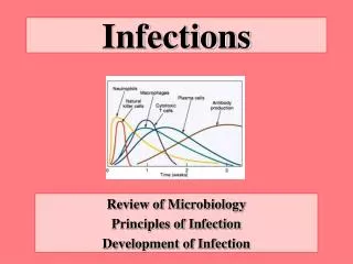
Viral • Epstein-BarrVirus: • EBV has developed into the ultimate B-lymphocyte parasite • EBV is a member of the subfamily Gammaherpesvirinae • The primary receptor for EBV is also the receptor for the C3d component of the complement system(also called CR2 or CD21). It is expressed on B cells of humans
EBV • causes heterophile antibody-positive infectious mononucleosisand • has been causally associated with • AfBL (endemic Burkitt lymphoma), • Hodgkin disease, and • nasopharyngeal carcinoma. • EBV has also been associated with B-cell lymphomas in patients with acquired or congenital immunodeficiencies. • EBV stimulates the growth and immortalizes B cells in tissue culture.
EBV • EBV encodes more than 70 proteins, different groups of which are expressed for the different types of infections. • The main viral proteins produced: • early antigen (EA), • viral capsid antigen (VCA), • Epstein-Barr nuclear antigens (EBNAs) • latent proteins (LPs); • latent membrane proteins (LMPs) 1 and 2; • and two small Epstein-Barr-encoded RNA (EBER) molecules, EBER-1 and EBER-2.
EBV • The diseases of EBV result from • either an overactive immune response (infectious mononucleosis) or • the lack of effective immune control (lymphoma and hairy cell leukoplakia)
Viral • Disease Mechanisms of Epstein-Barr Virus: • Virus in saliva initiates infection of oral epithelia and spreads to B cells in lymphatic tissue. • There is productive infection of epithelial and B cells. • Virus promotes growth of B cells (immortalizes). • T cells kill and limit B-cell outgrowth. T cells are required for controlling infection. Antibody role is limited.
Viral • Disease Mechanisms of Epstein-Barr Virus: • EBV establishes latency in memory B cells and is reactivated when the B cell is activated. • T-cell response (lymphocytosis) contributes to symptoms of infectious mononucleosis. • There is causative association with lymphoma in immunosuppressed people and African children living in malarial regions (African Burkitt lymphoma) and with nasopharyngeal carcinoma in China.
Infectious mononucleosis • results from a "civil war" between the EBV-infected B cells and the protective T cells. • The T cells are surrounded by infected B cells and are activated by viral antigenic peptides presented on both the MHC I and II molecules. The classic lymphocytosis (increase in mononuclear cells), swelling of lymphoid organs (lymph nodes, spleen, and liver), and malaise associated with infectious mononucleosis results mainly from the activation and proliferation of T cells.
Infectious mononucleosis • The T cells appear as atypical lymphocytes (also called Downey cells) • They increase in number in the peripheral blood during the second week of infection, accounting for 10% to 80% of the total white blood cell count at this time (hence the "mononucleosis"). • Children have a less active immune response to EBV infection and therefore have very mild disease.
Atypical T cell (Downey cell) characteristic of infectious mononucleosisThe cells have a more basophilic and vacuolated cytoplasm than normal lymphocytes, and the nucleus may be oval, kidney shaped, or lobulated. The cell margin may seem to be indented by neighboring red blood cells.
EBV • The virus persists in at least one memory B cell per milliliter of blood for the person's lifetime. • EBV may be reactivated when the memory B cell is activated (especially in the tonsils or oropharynx) and may be shed in saliva.
EBV • is transmitted in saliva . • More than 90% of EBV-infected people intermittently shed the virus for life, even when totally asymptomatic. • Children can acquire the virus at an early age by sharing contaminated drinking glasses. Children generally have subclinical disease
EBV • Saliva sharing between adolescents and young adults often occurs during kissing; thus EBV mononucleosis has earned the nickname "the kissing disease." • Disease in these people may go unnoticed or may manifest in varying degrees of severity. • At least 70% of the population of the United States is infected by age 30. • In developing countries more than %90
EBV • The geographic distribution of some EBV-associated neoplasms indicates a possible association with cofactors. • The immunosuppressive potential of malaria has been suggested as a cofactor in the progression of chronic or latent EBV infection to AfBL. • The restriction of nasopharyngeal carcinoma to people living in certain regions of China indicates a possible genetic predisposition to the cancer or the presence of cofactors in the food or environment. • More subtle mechanisms may facilitate the role of EBV in 30% to 50% of cases of Hodgkin disease
EBV • Transplant recipients, • patients with the acquired immune deficiency syndrome (AIDS), and genetically immunodeficient people are at high risk for lymphoproliferative disorders initiated by EBV. • These disorders may appear as polyclonal and monoclonal B-cell lymphomas. Such people are also at high risk for a productive EBV infection in the form of hairy oral leukoplakia.
Hairy oral leukoplakia Uncontrolled lytic infection by EBV
Diagnosis of EBV infection • By serology: • Heterophile antibody: • Recognition of Paul-Bunnell antigen on sheep, horse, or bovine erythrocytes • EBV-induced B-cell proliferation promotes production of heterophile antibody • Early symptom occurs in more than 50% of patients MONOTEST
If monotest is negative Ask for ELISA VCA-IgM
Diagnosis of EBV infection in • Transplant recipients, • patients with the acquired immune deficiency syndrome (AIDS), and genetically immunodeficient people to follow if lymphoproliferative disorders areinitiated by EBV: EBV DNA quantitation by real-time PCR
Retroviruses • Oncoviruses:immortalize or transform target cells, A,B,C,D type according to their core and capsid • Lentiviruses:slow viruses associated with neurologic and immunosuppresive disease • Spumaviruses:no disease • Endogenous viruses:transmitted vertically, 1% of human chromosome, in many animal species and humans, one detected in placental tissue which facilitates placental function
AIDS cause by HIV • Retroviridae family • Lentivirinae genus • Enveloped, positive strand RNA virus • 2 identical 9-10kb RNA
AIDS • Human immunodeficiency virus type 1 and 2 (HIV-1, HIV-2)
HIV • HIV-1: isolated in1983 • Responsible from AIDS pandemic • HIV-2: isolated in 1986 • HIV-2 less pathogenic slow progression to AIDS
HIV group and subtypes • Rapid mutation and recombination HIV-1 • Group M (major): A-J • Group O HIV-2 • A-E subtypes
Gp120 • CD4 surface receptor protein • Initially expressed on cells of the macrophage lineage (macrophage, dendritic cells, microglial cells) (M-tropic)+ second receptor CCR5 • Later on helper T cells (T-tropic) +fusin (CXCR4)
Disease Mechanisms of HIV • Human immunodeficiency virus primarily infects CD4 T cells and cells of the macrophage lineage (e.g., monocytes, macrophages, alveolar macrophages of the lung, dendritic cells of the skin, and microglial cells of the brain).
Disease Mechanisms of HIV • Virus causes lytic infection of CD4 T cells and persistent low-level productive infection of macrophage lineage cells. • Virus causes syncytia formation, with cells expressing large amounts of CD4 antigen (T cells); subsequent lysis of the cells occurs.
Disease Mechanisms of HIV • Virus alters T-cell and macrophage cell function. • Virus reduces CD4 T cell numbers and helper-cell maintenance of CD8 T cell and macrophage function. • CD8 T cell numbers and macrophage function decrease.
Acute retroviral syndrome • First signs occur in days to several weeks • Transient • %50-70 • Activation of immune system • Multisystem dysfunction • Flu or infectious mononucleosis sydrome like findings • Then a latent period
AIDS • Continuous viral replication • Immune system dysfunction
CD4 T lymphocyes • > 500 /µl (> %29) 1 • 200-499 (% 14-28) 2 • < 200 /µl (< %14) 3
Incubation • Adults with no treatment: 10-11 years • ‘rapid progressors’ : 2-3 years • ‘non-progressor’ : 7-10 years Stable CD4 cell count
AIDS may be manifested in several different ways • Lymphadenopathy and Fever • Opportunistic Infections • Malignancies • Dementia Related to AIDS
HIV • Viral RNA (in free Virion ) • Viral DNA: integrated in host cell DNA (Provirus)
Seroconversion • Usually 3 weeks • 1.5,3,6,12 months