Download
1 / 25
250 likes | 549 Views
Osseous Tissue & Bone Structure Ch 6. Coach Murray. Objectives. Classify bones according to shape & internal tissues Identify bone markings and significance of each type Identify the cell types in bone and their functions. Compare structure and function of compact bone and spongy bone

E N D
Osseous Tissue & Bone Structure Ch 6 Coach Murray
Objectives • Classify bones according to shape & internal tissues • Identify bone markings and significance of each type • Identify the cell types in bone and their functions. • Compare structure and function of compact bone and spongy bone • Compare the mechanisms of intramembranous ossification and endochondral ossification
Objectives • Describe the remodeling and homeostatic mechanisms of the skeletal system • Discuss the relationship of nutrition, hormones, exercise, and aging on bone development and on the skeletal system • Describe the types of fractures and explain how they heal
I. Skeletal System Functions pg 180 • Structural Support – a framework for attachment of soft tissues and organs • Storage of minerals – Ca, PO4 • Storage of lipids – in yellow marrow • Blood cell production – in red marrow • Protection – skull, vertebrae, pelvis, ribs • Leverage – change magnitude/direction of force of muscles
II. Gross Anatomy of Bones A. Six Categories of Bones by shape • Long – arms, bones, hands/feet, fingers/toes • Flat – roof of skull, sternum, ribs, scapula • Sutural (Wormian) – small, flat irregular in skull w/ borders like a jigsaw puzzle • Irregular – vertebrae, pelvis, some in skull • Short – small and boxy, tarsal and carpal bones • Sesamoid – sesame seed shaped, form near joints of knees, hands, and feet – i.e. patella
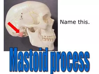
II. Gross Anatomy of Bones B. Bone Markings (Surface features of) – see Table 6-1 pg 182 • Elevations/Projections: process, ramus • Tendon/Ligament attachment: trochanter, tuberosity, tubercle, crest, line, spine • Articulation: head, neck, condyle, trochlea, facet • Depressions: fossa, sulcus • Openings: foramen, canal, fissure, sinus/antrum
II. Gross Anatomy of Bones C. Bone Structure pg 183 – of long bone Diaphysis – extended/expanded tubular shaft w/wall of dense/compact bone (cortex) Epiphysis – at the ends of diaphysis, consists largely of cancellous (spongy )bone Metaphysis – narrow zone b/w each epiphysis and the diaphysis
II. Gross Anatomy of Bones C. Bone Structure pg 183 (continued) 1. Long bone – a diaphysis (shaft) with an epiphysis at each end w/a metaphysis b/w them. Compact bone surrounds marrow cavity (medullary) and epiphyses are made cancellous bone (spongy). Note: Spongy bone is an open network of struts and plates with a cortex of compact bone. 2. Flat bone Compact bone surrounds diploe (spongy), marrow present, no marrow cavity
III. Bone Histology • Bone is - Osseous tissue is a supporting connective tissue with a solid matrix and ensheathed by a periosteum. • A. Bone Matrix basics: • Osteocytes: mature bone cells that maintain matrix • Lacunae: pockets which house osteocytes • Canaliculi: passageways through matrix • Periosteum: superficial layer of compact bone w/fibrous outer layer and cellular inner layer
III. Bone Histology • B. The Matrix of Bone: - pg 184 • Ca3(PO4)2 – 66% by mass • Are hard, inflexible, withstand compression • Interacts w/ Ca(OH)2 to make crystals of hydroxyapatite • Hydroxyapatite also contains CaCO3, Na, Mg, F • Collagen fibers – 31% by mass • Flexible, twist, bend, do not compress • Provide framework on which hydroxyapatite crystals form • Stronger than steel w/subject to tension (pull) • Cells of bone – 2%
III. Bone Histology • C. The Cells of Bone - structure and function of each • 4 kinds of cells 1) Osteocyte • Structure: housed in a lacuna sandwhiched b/w layers called lamellae. Canaliculi connect lamellae w/each other and central canal • Functions (2): • Maintain protein and mineral content of matrix • Repair damaged bone by converting to osteoblasts or osteoprogenitor cells
III. Bone Histology • C. Cells of Bone - structure and function of each • 4 kinds of cells 2) Osteblasts • Structure: immature osteocyte • Function: perform osteogenesis by converting osteoid to bone. Make and releasing proteins and other organic compounds into matrix, once surrounded they become osteocytes. This is bone building • Activity is constant, is increased w/exercise
III. Bone Histology • C. The Cells of Bone - structure and function of each • 4 kinds of cells 3) Osteoprogentior cells • Structure: type of stem cells which make osteoblasts. Found in 3 places: • Cellular layer of periosteum • endosteum (marrow cavities lining) • Lining of passageways containing blood vessels • Function: • Repair fractures/breaks in bone
III. Bone Histology • C. The Cells of Bone - structure and function of each • 4 kinds of cells 4) Osteoclasts (not related osteoprogenitor cells) • Structure: big multinucleated cells • Function: osteolysis - secrete acid and proteolytic enzymes to dissolve the matrix and release stored minerals. Regulates Ca & PO4 in body fluids • Opposite to activity of osteoblasts
D. Structure of Compact Bone pg 185 • Structure Described: • Haversian system (or osteon) is basic functional unit of compact bone arranged in concetric layers around the Haversion canal (or central canal) - • Structure: house blood vessels, run parallel to bone surface • Function: supply blood to osteons Other Info: Canals of Volkmann (perforating canals) – • Structure: house blood vessels, run perpendicular to bone surface • Function: supply blood to osteons of deeper in bone and marrow cavity
D. Structure of Compact Bone pg 185 • Structure Described: • So the lamellae of each osteon forms nested cylinders around central canal • 3 kinds of lamellae • Concentric lamellae – surround central canal, collagen forms a spiral, canaliculi interconnect lacunae of the osteon w/one another and w/the central canal • Interstitial lamellae – space b/w osteons, being recycled by osteoclasts • Circumferential lamellae – outer and inner surfaces of bone so they are covered in periosteum and endosteum
E. Structure of Spongy Bone pg 185 • Unlike compact bone, lamellae present but is not arranged in osteons • Matrix (description of): • Made of trabeculae – branched, open network of thin struts and plates • No capillaries/venules, nutrients get to osteocytes via diffusion along canaliculi openings • Red marrow b/w trabeculae has blood vessels to deliver nutrients and remove osteocyte wastes
F. Structure of Spongy Bone pg 185 • Located of little stress or where stress is multidirectional • Is lighter than compact, makes skeleton lighter • 2 kinds of marrow: • Red bone marrow: • Location: epiphyses of long bones, interior of ilium/sternum • Function: makes red blood cells • Yellow • Location: • Function:
G. The Periosteum and Endosteum pg 188 • Periosteum • Location: wraps superficial layer of compact bones except at joints • Structure: outer layer is fibrous, inner is cellular • Collagen fibers are continous w/bone, joint capsules, and attached tendons and ligaments providing stronger attachment • Functions: • 1) isolates bone from surrounding tissues • 2) provides route for circulatory/nervous supply • 3) participates in bone growth/repair
G. The Periosteum and Endosteum pg 188 • Endosteum • – location: lines marrow cavity, covers the trabeculae of spongy bone, lines inner surfaces of central canals • - functions: • 1) aid in growth, repair, remodeling • structure: single, incomplete layer of osteoprogenitor cells that cover the bone matrix • Exposed areas osteoclasts and osteoblasts can remove/deposit matrix
IV. Bone Formation and Growth pg 189 • Ossification – replacing tissues with bone during bone formation • Calcification – deposition of calcium salts which occurs during ossification • 2 kinds of ossification • A. Endochondral ossification • B. Intramembranous ossification
IV. Bone Formation and Growth pg 189 • A. Endochondral ossification – bone replaces existing cartilage • B. Intramembranous ossification – bone develops from mesenchyme or fibrous connective tissue • C. The Blood and Nerve Supplies • 3 major sets and where they provide blood to: • 1. nutrient artery and vein - diaphysis • 2. metaphyseal vessels – ephiphyseal cartilage • 3. periosteal vessels – superficial osteons of shaft
V. Dynamic Nature of Bone pg 194 • Remodeling – is bone maintenance – is the recycling and renewal of organic and mineral compounds of bone matrix • Requirements for osteogenesis: • Minerals in diet : Ca, PO4 , Mg, F, Fe, others • Vitamins: A, C, B12, K • Hormones: Calcitonin and parathyroid hormone
Resources • http://www.google.com/imgres?num=10&um=1&hl=en&safe=active&biw=1024&bih=599&tbm=isch&tbnid=ZugqcMvLVMfSlM:&imgrefurl=http://www.rci.rutgers.edu/~uzwiak/AnatPhys/APFallLect8.html&docid=75prWH10pRBdpM&imgurl=http://www.rci.rutgers.edu/~uzwiak/AnatPhys/APFallLect8_files/image003.jpg&w=640&h=480&ei=6Pt7ULvGF86u2AXWyIDYDQ&zoom=1&iact=hc&vpx=490&vpy=190&dur=159&hovh=194&hovw=259&tx=159&ty=121&sig=112719699817004934008&sqi=2&page=2&tbnh=140&tbnw=187&start=14&ndsp=16&ved=1t:429,r:16,s:0,i:116