Download
1 / 75

E N D
GramStaining in Clinical Microbiology Dr.T.V.Rao. MD Professor of Microbiology Travancore Medical College, Kollam. Kerala Dr.T.V.Rao MD
The Guest Lecture presented by Dr.T.V.Rao MD at • Tenth National Workshop on • “Simple Diagnostic Methods in Clinical Microbiology” • 29th November to 3rd December 2011 • Department of Microbiology • Jawaharlal Institute of Postgraduate Medical Education & Research, Pondicherry Dr.T.V.Rao MD
Working at Mansa General Hospital Mansa Republic of ZambiaTaught many lessons, to understand Infection and Ignorance are important causes of Morbidity and Mortality Dr.T.V.Rao MD
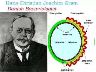
Hans Christian Gram • The Gram stain was devised by the Danish physician, Hans Christian Gram, while working in Berlin in 1883. He later published this procedure in 1884. At the time, Dr. Gram was studying lung tissue sections from patients who had died of pneumonia. Dr.T.V.Rao MD
First Paper on Gram Staining • In his paper, Dr. Gram described how he was able to visualize what we now call Staphylococcus, Streptococcus, Bacillus, and Clostridiain various histological sections. Interestingly, Dr. Gram did not actually use safranin as a counter stain in the original procedure (Gram negative cells would be colorless). He instead recommended using Bismarck brown as a counter stain to enable tissue cell nuclei to be visualized. Dr.T.V.Rao MD
Carl Weigert (1845-1904) • German pathologist Carl Weigert (1845-1904) from Frankfurt, added a final step of staining with safranin. Dr.T.V.Rao MD
Traditional Definition of Gram stain • A method of staining bacteria using a violet stain. The gram staining characteristics (denoted as positive or negative). A heat fixed bacterial smear is stained with crystal violet (methyl violet), treated with 3% iodine/potassium iodide solution, washed with alcohol and counterstained. The method differentiates bacteria into two main classes, gram-positive and gram-negative. Dr.T.V.Rao MD
The Cell walls differ… Dr.T.V.Rao MD
Gram Positive should not be Mistaken • In the Gram Stain technique, two positively charged dyes are used: crystal violet and safranin. The use of the designation “gram-positive” should not be confused with the concept of staining cells with a simple stain that has a positive charge. Dr.T.V.Rao MD
Gram staining observation Basic Principle in Koch’s postulations • The first of Koch’s postulatethat the suspected the organism should always be found in association with the disease. Dr.T.V.Rao MD
Poor quality of slidesCan be corrected • Use of glass slides that have not been pre cleaned or degreased ? NOTE: Storing slides in a jar with 95% ethanol will ensure clean slides. Drain excess alcohol or flame slide before use. Dr.T.V.Rao MD
Four Major Steps in Gram Staining • There are four basic steps of the Gram stain, which include applying a primary stain (crystal violet)or Methyl violet to a heat-fixed smear of a bacterial culture, followed by the addition of a mordant (Gram's iodine), rapid decolorization with alcohol or acetone, and counterstaining with Safranin or basic fuchsin. Dr.T.V.Rao MD
Organizing the Staining Bottles Dr.T.V.Rao MD
Making a Smear • First prepare your slide. You do this by placing bacteria on a slide in a drop of water, allowing them to dry and then heat fixing them. Heating Dr.T.V.Rao MD
Correct preparation • Smear preparation: Proper smear preparation should produce a monolayer of organisms sufficiently dense for easy visualization but thin enough to reveal characteristic morphological characteristics. Use clean, new glass slides. NOTE: When using the same pipette or swab, always inoculate culture media first Dr.T.V.Rao MD
Method of smearing the MaterialWrong Right Dr.T.V.Rao MD
Using Methanol is it Better than Heat Fixation ? • Fix the smear with 95% Methanol • Which will help in prevention of distortion of cells • Helpful in Microscopic observation of CSF and Urine Dr.T.V.Rao MD
Making Multiple smears in same slide – conserve resources • Making multiple smears make the optimal use of the slide. • Reduces the economic costs and saves the technical time. Dr.T.V.Rao MD
Steps in Gram Staining Procedure- Follow the Clock • 1On a rack, flood with filtered crystal violet ( Methyl violet ) 10 sec 2 Wash briefly in water to remove excess crystal violet • 3. Flood with Gram’s iodine 10 sec • 4. Wash briefly in water, do not let the section dry out. • 5. Decolourise with acetone for few seconds <6 seconds until the moving dye front has passed the lower edge of the section • 6. Wash immediately in tap water • 7. Counterstain with safranin for 15 seconds.. Dr.T.V.Rao MD
Proceed in organized Fashion Dr.T.V.Rao MD
Step 1 Dr.T.V.Rao MD
Step 2 Dr.T.V.Rao MD
Step 3 Dr.T.V.Rao MD
Step 4 Dr.T.V.Rao MD
Step 5 Dr.T.V.Rao MD
How long you keep Iodine in the Laboratory ??? • The Gram’s Iodine we make in the laboratory from basic chemicals • How long we can use it ? • Why we have to make frequently ? Dr.T.V.Rao MD
Most Critical Step in Gram staining • The most critical step of gram staining is the decolorization step as crystal violet stain will be removed from both G+ve & G-ve cells if the decolorizing agent(e.g alcohol ) is left on too long. Dr.T.V.Rao MD
Acetone used with Caution • Acetone is a more rapid decolorizes than alcohol and must be used with some care. • Excessive decolorization turns Gram positive appear as Gram negative Dr.T.V.Rao MD
Which alcohol is better • Several alcohols have been studied, and it has been reported that the more complex the alcohol, the slower the decolorization action. As the carbon chain lengthens, decolorization is slower. Conn found in practice, however, no known advantage can be gained by substituting the higher alcohols for ethyl alcohol. Dr.T.V.Rao MD
Step 6 Dr.T.V.Rao MD
Which counterstain is better • Some bacteria which are poorly stained by Safranin,such as Hemophilus spp., Legionella spp., and some anaerobic bacteria, are readily stained by basic fuchsin, but not Safranin Dr.T.V.Rao MD
Step 7 Dr.T.V.Rao MD
Caring the stained slide After the counterstain has been rinsed off, the slide is placed between some absorbent paper and the excess water gently blotted off. Care must be taken not to rub the slide with the blotting paper because this would remove the adhering bacteria. Dr.T.V.Rao MD
Gram staining depends on • Includes culture age, media, incubation atmosphere, staining methods, . Similar considerations apply to the interpretation of smears from clinical specimens, and additional factors include different host cell types and possible phagocytosis. • Gram stain permits the separation of all bacteria into two large groups Dr.T.V.Rao MD
How the Gram Stain Work • So how does it work? Gram didn't know - he simply worked empirically. We now know that the Gram reaction is based on the structure of the bacterial cell wall. • In Gram-positive bacteria, the dark purple crystal violet stain is retained by the thick layer of peptidoglycan which forms the outer layer of the cell. • In Gram-negative bacteria, the thin peptidoglycan layer in the periplasm does not retain the dark stain, and the pink safranincounterstains the peptidoglycan layer. Dr.T.V.Rao MD
Optimal use of Microscopy • Gram stained preparations have to be observed with bright-field optics. Phase-contrast microscopy does not allow the recognition of true colours. Gram-positive bacteria may be seen under phase-contrast as red cells. Using bright-field optics, Gram-positive cells are purple or blue and Gram-negative pinkdue to counter stain with Safranin.. Dr.T.V.Rao MD
Report as follows • 1 If no microorganisms are seen in a smear of a clinical specimen, report “No microorganisms seen.” • 2. If microorganisms are seen, report relative numbers and Describe morphology. • Observe predominant shapes of microorganisms Dr.T.V.Rao MD
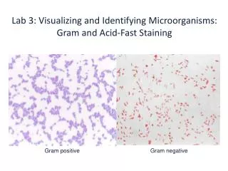
A gram stained bacterial suspension containing a mixture of Gram negative bacilli, and Gram positive cocci arranged in bunches (Staphylococci spp)
A true Gram Negative staining Dr.T.V.Rao MD
Value of Direct Smears • Guide the physician on initial choice of antibiotic, pending results of culture and sensitivity. • Judge specimen quality. • Contribute to selection of culture media, especially with mixed flora. • Provide internal quality control when direct smear results are compared to culture results. Dr.T.V.Rao MD
Staining depends on Staining depends on Structural Integrity of Cell Wall • We know that only intact cells are Gram-positive, so that cells which are even gently broken become Gram-negative. Observations suggest that bacterial protoplasts, devoid of cell wall, are still Gram-positive, indicating that it is probably the semipermeable membrane which is somehow involved in the reaction. Dr.T.V.Rao MD
Nature of Morphology guides early Diagnosis in uncommon diseases Dr.T.V.Rao MD
Identify • A young patient presented with foul smelling purulent discharge since 2 days on observation by Gram staining
Observe Spores may appear as Gram negative and Gram positive
Burkholderia pseudomallei is a gram-negative bacilli with a “safetypin” appearance on microscopic examination Dr.T.V.Rao MD
Limitations of Gram’s Staining • We know that Gram positivityis restricted almost exclusively to the bacteria, with only a few other groups, such as the yeasts, exhibiting this reaction. Dr.T.V.Rao MD
Better Understanding of Gram’s Staining • We should know that the Gram stain is not an all-or-nothing phenomenon, but that quantitative variations in Gram-positivity exist between different species,and within the same species during different parts of the growth cycle or under different environmental conditions. Dr.T.V.Rao MD
Stains Several Fungi Dr.T.V.Rao MD
Streptococcus pneumonia Dr.T.V.Rao MD