Download
1 / 34
420 likes | 1.85k Views
Management of Patients Receiving Radiation Therapy. Objectives. The learner will be able to: Identify the various types of radiation therapy used to treat cancer. List education priorities for the patient receiving radiation therapy. Radiation Safety . Cardinal principles

E N D
Objectives The learner will be able to: • Identify the various types of radiation therapy used to treat cancer. • List education priorities for the patient receiving radiation therapy.
Radiation Safety • Cardinal principles • Decrease time of exposure to radiation. • Increase the distance from radiation exposure. • Use shielding devices to absorb radiation. • Radiation monitoring devices • Individual personal dosimetry badge: • Ring dosimeter: worn by person handling radioactive material
Radiation-Restricted Areas • Universal radiation caution signs are posted in any area with potential or actual radiation exposure. • Radiation safety precautions are posted for any patient receiving radionuclide therapy. • Radiation safety sheets are posted in patient’s chart.
Special Populations • Pregnant women • Most institutions recommend that pregnant women not care for patients receiving radionuclide therapy • Employee assumes all responsibility for exposure of the fetus until pregnancy is officially declared.
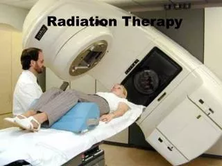
External Beam Radiation Therapy (EBRT) • EBRT is the most common form of radiotherapy. • Radiation is delivered from outside the body. • Linear accelerator is the most common treatment machine used to deliver EBRT.
EBRT: Indications • Can be the primary treatment • Used before surgery to shrink tumor • Used after chemotherapy or surgery to get tumor cells left behind • Delivered to high-risk areas to prevent cancer growth • Used to control cancer • Used to manage symptoms or to improve quality of life • Used to treat structural emergencies
EBRT: Pretreatment • Imaging and staging diagnostics • Blood tests or cancer markers • Evaluation by radiation oncologist and/or medical oncologist as indicated
EBRT: Treatment Planning • Simulation • Obtain images for treatment planning. • Immobilization devices are made. • Determine treatment position each day. • Treatment planning • Based on CT, MRI, and PET/CT scans • Determine volume of tumor to be treated. • Computer calculation of dose to tumor and surrounding tissues
EBRT: Treatment Delivery • Once a day, five days/week (MonFri) • Treated two to nine weeks based on tumor type • Actual beam is on for few minutes, with the rest of time used for positioning. • Patient does not feel anything during treatment. • Patient is not radioactive after treatment.
EBRT: Side Effects • Side effects are specific to area treated. • Fatigue is usually experienced despite area treated and increases as treatment proceeds. • Side effects usually occur during treatment and resolve in two weeks. • Based on area treated, some patients will have long-term side effects.
EBRT: Post-Radiation Care • Periodic follow-up treatment visits with the physician • Imaging studies to see if the cancer has responded to treatment • Blood test and tumor markers • Physical exam and evaluation
EBRT: Patient Education • Radiation is a local/regional treatment. • Concurrent therapy (RT and chemotherapy) to optimize treatment outcomes • Side effects are specific to area treated. • Side effects usually occur during treatment and resolve in two weeks. • Some patients will experience long-term side effects. • Your doctor or nurse will work with you to manage your side effects.
Brachytherapy • Indications • Temporary or permanent placement of a radioactive source into: • Body cavity (intracavitary) • Tissue (interstitial) • On the surface of the body • Can be used in conjunction with EBRT • Two types of brachytherapy • Low dose rate (LDR) • High dose rate (HDR) • May be used as a “boost” with EBRT
Brachytherapy: Goals of Treatment • Improve local tumor control. • Irradiate small volumes. • Potentially minimize complications. • Preserve organ function. • Treat recurrent or inoperable cancers. • Control disease in previously irradiated sites.
LDR Brachytherapy • Hospitalized: Operative procedure with anesthesia • Hollow applicator device or catheter is placed. • Radioactive sources are manually loaded once patient returns to room. • Strict room confinement • Bed rest required for gyn, rectal and some prostate implants • Specialized nursing care in hospital • Requires radiation precautions
HDR Brachytherapy • Involves the use of automated remote afterloading devices for placement of the radioactive source • HDR treatments done as an outpatient • Treated with high doses of radiation in shorter treatment times ( but more treatments may be needed) • Anesthesia or sedation may be required depending on the site, applicator, and age or comprehension of the patient
Brachytherapy: Pretreatment • LDR • Pretreatment bowel regimen (enema) on morning of procedure • Educate patient on respiratory complications and prevention of immobility. • Anticoagulation if indicated • HDR • Foley catheter and rectal tube may be placed. • Premedicate with pain and anxiety medications. • Radiation implant briefs for gynimplants
Brachytherapy: Post-Radiation Care • LDR • Antidiarrheals to minimize bowel movements • Low-residue diet (with finger foods) • HOB not elevated more than 30 degrees • Modify bathing and linen changes • Prevent complications of immobility • Deep breathing/coughing reinforcement • Use of compression stockings • Isometric exercises • Anticoagulants as ordered
Brachytherapy: Side Effects • Localized and involves only the site implanted • Pain and swelling of tissue implanted • Soft tissue injury or necrosis (long-term) • Diarrhea, proctitis, nausea, moist desquamation in skin folds • GU symptoms • Side effects are managed using standard treatment strategies.
Brachytherapy: Reportable Signs/Symptoms • Excessive bleeding from site • Temp > 101°F • Drainage or foul discharge • Urinary symptoms • Diarrhea or constipation • Increased pain
Brachytherapy: Patient Education • Prepare patient for procedure and what is expected. • Prepare patient for the social isolation associated with strict radiation precautions. • Teach symptoms to report during treatment. • Explain the importance of prevention measures during immobilization (CDB, compression stockings)
Brachytherapy: Emergency Procedures • Dislodged sources • Notify radiation safety officer (RSO) immediately for dislodged sources. • Never pick up a dislodged source. • Use long-handle forceps to pick up source and place in lead container in room. • RSO scans everything before removing the source from the room.
Cardiopulmonary Resuscitation of patient who has received radionuclide therapy • Begin CPR immediately. • Wear gloves, gowns, and shoe covers. • Notify RSO immediately to remove sealed source and place in lead container. • All equipment should be scanned for radiation contamination before removal from the room. • All personnel performing CPR must be cleared by RSO before leaving the room.
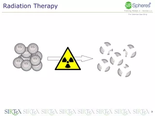
Radioisotopes and Radiopharmaceuticals • Radioisotopes • Used for palliation of bone pain • Generally administered via IV as outpatient • Can be used alone or in combination with bisphosphonates • Radiopharmaceuticals • Unsealed sources that can be ingested, injected, or instilled • Example: I-131 is used to treat thyroid cancer. • Sr-89 and Sm-153 are used to treat multiple bone metastases.
Radiopharmaceuticals • Very effective in treating specific tumors and have very few side effects • Dose • Less than 33 mCi: outpatient • Greater than 33 mCi: inpatient • Follow radiation precautions. • Follow body fluid precautions.
Radioisotopes and Radiopharmaceuticals: Side Effects • Leukocytopenia and thrombocytopenia may occur. • Bone pain flare • Erythema, tenderness or dryness of the skin
Radioisotopes and Radiopharmaceuticals: Assessment • Check blood counts one week prior to administration of radioisotopes. • Routine assessment of pain and effectiveness of pain regimen • Patient needs to continue taking analgesics (if treating bone pain); may take two to three weeks for response. • IV access if indicated
Radioisotopes and Radiopharmaceuticals:Management of Side Effects • Obtain blood counts one week after administration. • Aggressive pain regimen • Use of NSAIDs, opiates, and steroids for management of bone pain flare
Radioisotopes and Radiopharmaceuticals: Patient Education • Bone pain flare may occur 72 hours after administration and last up to one week. • Blood counts can be weekly for up to eight weeks. • Precautions should be taken for at least 12 hours after administration. • Flush toilet at least two times after each use. • Wash hands with soap and water after toileting. • Wash linens separately if exposed to body fluids.
References Eggert, J. (2010). Cancer basics. Pittsburgh, PA: Oncology Nursing Society. Iwamoto, R., Haas, M., & Gosselin, T. (2012). Manual for radiation oncology nursing practice and education (4th ed.). Pittsburgh, PA: Oncology Nursing Society.