Download
1 / 17
170 likes | 209 Views
Learn about aspergillosis, a disease caused by Aspergillus fungi, with its various types and clinical manifestations. Explore the diagnostic methods, including culture and histopathology, and understand the treatment options available.

E N D
ASPERGILLOSIS Prof. Khaled H. Abu-Elteen
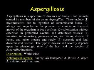
ASPERGILLOSIS Aspergilli produce a wide variety of diseases. Like the zygomycetes, they are ubiquitous in nature and play a significant role in the degradation of plant material as in composting. Similar to Candida and the Zygomycetes, they rarely infect a normal host. The organism is distributed world-wide and is commonly found in soil, food, paint, air vents. They can even grow in disinfectant. There are more than one hundred species of aspergilli. The most common etiologic agents of aspergillosis : Aspergillus fumigatus A. niger A. flavus
Aspergillus Conidia Conidiogenous cells (phialides) Supporting cell (branch or metulae) Swollen apex of conidiophore (vesicle) Conidiophore Basal part of conidiophore (foot cell)
Anamorphs--Penicillium phialides Branches (metulae)
Penicillium Aspergillus
There are three clinical types of pulmonary aspergillosis: • Allergic - hypersensitivity to the organism. Symptoms may vary from mild respiratory distress to alveolar fibrosis. • Aggressive tissue invasion. Primarily a pulmonary disease, but the aspergilli may disseminate to any organ. They may cause endocarditis, osteomyelitis, otomycosis and cutaneous lesions. • Fungus ball or Pulmonary Aspergilloma which is characteristically seen in the old cavities of TB patients. This is easily recognized by x-ray, because the lesion (actually a colony of mold growing in the cavity) is shaped like a half-moon (semi-lunar growth). The patients may cough up the fungus elements because the organism frequently invades the bronchus. Chains of conidia can sometimes be seen in the sputum.
Culture: Aspergilli require 1-3 weeks for growth. The colony begins as a dense white mycelium which later assumes a variety of colors, according to species, based on the color of the conidia. The hyphae are branching and septate. Species differentiation is based on the formation of spores as well as their color, shape and texture. Histopathology: The septate hyphae are wide and form dichotomous branching, i.e., a single hypha branches into two even hyphae, and then the mycelium continues branching in this fashion
Serology: There is an excellent serological test for aspergillosis which is an Immunodiffusion test ( ELISA). There may be 1 to 5 precipitin bands. Three or more bands usually indicate increasingly severity of the disease. i.e., tissue invasion.( detection of IgG and IgE) Treatment: Voriconazole, Fluconazole, Itraconazole and Amphotericin B.